13054_2025_Article_5392
Visão geral


Maia et al. Critical Care (2025) 29:155 Critical Care https://doi.org/10.1186/s13054-025-05392-w
RESEARCH Open Access
Peri‑intubation adverse events and clinical

outcomes in emergency department patients: the BARCO study
Ian Ward A. Maia1,2*, Bruno A. M. Pinheiro Besen3, Lucas Oliveira J. e Silva2,4, Rafael von Hellmann5,
Ludhmila Abrahao Hajjar1, Benjamin J. Sandefur2, Daniel Fontana Pedrollo4, Caio Goncalves Nogueira6, Natalia Mansur P. Figueiredo6, Carlos Henrique Miranda7, Danilo Martins8, Thiago Dias Baumgratz8, Bruno Bergesch9, Diogo Costa9, Osmar Colleoni10, Juliana Zanettini11, Ana Paula Freitas11,
Nicole Pinheiro Moreira12, Patricia Lopes Gaspar12,13, Renato Tambelli14, Maria Cristina Costa15,
Samara Silveira15, Wilsterman Correia16, Rafael Garcia de Maria17, Ubirajara A. Vinholes Filho18,
Andre P. Weber19, Vinicius da Silva Castro20, Carlos Fernando D. Dornelles21, Barbara S. Tabach21,
Hélio P. Guimarães1, Gabriela Stanzani1, Thiago F. Gava1, Aidan Mullan2, Heraldo P. Souza1,
Otavio T. Ranzani22,23, Fernanda Bellolio2, Julio C. G. Alencar24, on behalf BARCO group, Victor Paro da Cunha,
Julio F. Marchini, Patricia Albuquerque Moura, Fernanda Greco, Yasmine Filippo, Rubens Yoshinori Kai,
Guilherme Torres Abi Ramia Chimelli, Juan Valdivia, Edson Luiz Favero Junior, Felipe Rischini,
Vitor Amorim de Andrade Câmara, Henrique Bertotto, Victor Borges, Juliano Rathke, Renato Melo,
Ariadine Augusta Maiante, Sarah Maciel Silva, Clarisse Moreira Ribeiro de Oliveira, Andressa Pi Rocha Reis,
Thamyres de Carvalho Rufato, Gabriella Dias, Victoria Sartor Poloni, Kauê Lima, Hilana Zenly, José Carlos Motta,
Gabriel Miranda, Alexandre Freitas, Leonardo Gasperini, Thais Raimondi Sudbrack, Ana Paula Ribeiro,
Guilherme Henrique A. do Carmo, Andrea de Vargas Tomelero, Augusto Lengler Konrath,
Vitor Cremonese Zanella, Natalia Fuhr, Davi Amaral Cesário Rosa, Isabela Lopes Lima, Luiz Fernando Varela,
Isabella Baldino, Andre Zimmerman, Julia M. Dorn de Carvalho and Molly M. Jeffrey
Ian Ward A. Maia
ian.ward.maia@gmail.com
Full list of author information is available at the end of the article
© The Author(s) 2025. Open Access This article is licensed under a Creative Commons Attribution-NonCommercial-NoDerivatives 4.0 International License, which permits any non-commercial use, sharing, distribution and reproduction in any medium or format, as long as you give appropriate credit to the original author(s) and the source, provide a link to the Creative Commons licence, and indicate if you modified the licensed material. You do not have permission under this licence to share adapted material derived from this article or parts of it. The images or other third party material in this article are included in the article’s Creative Commons licence, unless indicated otherwise in a credit line to the material. If material is not included in the article’s Creative Commons licence and your intended use is not permitted by statutory regulation or exceeds the permitted use, you will need to obtain permission directly from the copyright holder. To view a copy of this licence, visit .
Maia et al. Critical Care (2025) 29:155 Page 2 of 15
Abstract
Background Emergency tracheal intubation in critically ill patients carries a high risk of complications, and practices vary substantially across different settings. Identifying risk factors and understanding how peri-intubation adverse events affect patient outcomes may guide standardization of care and improve survival.
Methods This prospective cohort study involved 18 emergency departments in Brazil (March 2022–April 2024). We included adults (≥ 18 years) undergoing emergency intubation and excluded patients intubated electively or for car-diac arrest. We defined major peri-intubation adverse events as severe hypoxemia, new hemodynamic instability, or cardiac arrest occurring within 30 min of initiating intubation. The primary outcome was 28-day mortality. Multi-variable regression analyses assessed associations between adverse events and mortality, controlling for potential confounders.
Results Among 2846 patients, major adverse events occurred in 919 (32.3%) intubations, most frequently new hemodynamic instability (20.0%), followed by severe hypoxemia (12.5%) and cardiac arrest (3.5%). The overall 28-day mortality was 45.1%. Patients experiencing any major adverse event had a significantly higher 28-day mortality (57.6 vs 39.2%; aHR 1.43, 95% CI 1.26–1.62; p < 0.001). Sensitivity analyses confirmed these findings. Successful first-attempt intubation was associated with a reduced likelihood of major adverse events (aOR 0.52; 95% CI 0.41–0.65; p < 0.001).
Conclusion One in three patients undergoing emergency intubation experienced a major peri-intubation adverse event, which was associated with higher 28-day mortality. These results underscore the importance of optimizing intubation strategies to reduce complications and potentially improve patient outcomes in critically ill patients.
Keywords Intubation, Airway registry, Difficult airway, Adverse events, Critical illness, Emergency airway management
Background
Tracheal intubation (TI) is a critical procedure per-formed in emergency departments (EDs) and intensive care units (ICUs) worldwide [1]. While essential for man-aging critically ill patients, it carries significant risks, par-ticularly in resource-limited settings [1–3]. Major adverse events (MAEs), such as hemodynamic instability, severe hypoxemia, and cardiac arrest, are frequently observed in the peri-intubation period and have been associated with worse patient outcomes [3–6].
Critically ill patients frequently present with a ‘physi-ologically difficult airway,’ characterized by acute hemo-dynamic instability, compromised oxygenation, and metabolic disturbances that increase the risk of peri-intubation adverse events, even in the absence of ana-tomical airway difficulty [7, 8]. An international cohort study involving 29 countries found that the incidence of MAEs during emergency intubations exceeded 40% [4]. The study reported a first-attempt intubation success rate of nearly 80%, with only 4.5% of patients requiring more than two attempts. This high incidence of MAEs, despite relatively high first-attempt success, suggests that factors beyond anatomic challenges contribute to adverse events [4, 9–11].
Understanding potential causal pathways in airway management is crucial for improving outcomes in criti-cally ill patients undergoing emergency intubations globally [12–14]. However, the global burden of these complications remains poorly understood in low- and
middle-income countries (LMICs), since prospective studies examining the association between peri-intu-bation adverse events and 28-day mortality are limited [4–6].
Therefore, we established the Brazilian Airway Registry Cooperation (BARCO), the first multicenter registry of emergency intubations in a middle-income country. This study aims to determine the incidence of MAEs and their association with 28-day mortality in critically ill patients undergoing emergency intubations, offering essential data from a resource-limited setting.
Methods
Study design and setting
We conducted a prospective cohort study across 18 EDs in Brazil, spanning four regions, as part of the BARCO network. We report these results in accordance with the Strengthening the Reporting of Observational Studies in Epidemiology (STROBE) statement [15]. This study was approved by the ethics committees of all participat-ing centres. Due to the observational nature of the study and the use of de-identified data, a waiver of individual patient consent was granted by each institution’s ethics committee.
Participants
We enrolled adults (age ≥ 18 years) undergoing TI in the ED. Exclusion criteria were intubations performed for elective procedures or during cardiac arrest. Tracheal
Maia et al. Critical Care (2025) 29:155 Page 3 of 15
intubations were performed by clinical staff working in the ED. Specific procedural aspects, including the choice of medications, equipment, and intubation techniques, were determined according to clinicians preferences and standard practices at each participating center. To ensure comprehensive data collection and avoid selective report-ing, each site investigator submitted a study compliance plan approved by the coordinating center (Hospital das Clínicas de São Paulo, SP, Brazil). This plan detailed the process for identifying consecutive ED intubations and ensuring at least 80% were recorded in the BARCO data-base. Monthly compliance reports were submitted and reviewed by BARCO coordinators for quality control.
Exposures and outcomes
Our primary exposures were MAEs, defined as the pres-ence of one or more of the following within 30 min from the start of the intubation procedure: severe hypoxemia (peripheral oxygen saturation < 80%), new hemodynamic instability (systolic arterial pressure < 65 mmHg recorded at least once, new requirement for or increase the dose of vasopressors, or administration of a fluid bolus > 15 mL/ kg to maintain target blood pressure), or cardiac arrest [4]. Our primary outcome was 28-day mortality after TI, assessed through electronic health record reviews, with follow-up until hospital discharge, death, or 28 days, whichever occurred first. Secondary outcomes included the incidence of MAEs, difficult intubations (defined as three or more attempts), the first-attempt success, tran-sient hypotension, defined as a single episode of systolic blood pressure < 90 mmHg or mean arterial pressure < 65 mmHg that does not fulfill the criteria for new hemody-namic instability, and esophageal intubation.
Data collection
data collection form using a survey on REDCap®. [16, 17] We required the form to be completed within 30 min of tracheal intubation confirmation. A site investigator at each center trained staff on completing the form and des-ignated a trained observer rather than the clinician who performed the intubation to complete the form.
We collected variables representing patient charac-teristics, illness severity, preprocedural physiology, and intubation characteristics. Definitions for all collected variables are available in eMethods1. (Appendix File).
Statistical analysis
As this study was designed to be a prospective observa-tional airway management registry, we did not calculate a target sample size. Given the inclusion of 18 centers with a minimum of 40 intubations to be included, the minimum sample size would be 720 intubations, but
we estimated that 3–5 times more participants would be included given the center’s volumes of inclusion, which would allow sufficient power to estimate the rate of adverse events, 28-day mortality and to explore their association.
We present descriptive statistics as mean ± SD or median (interquartile range [IQR]) for continuous vari-ables, frequency and proportion for categorical variables, stratified by peri-intubation MAEs. We performed bivar-iate analyses with χ2 or Fisher’s exact test for categorical variables, and the Mann–Whitney or t-test for continu-ous variables, as appropriate. We plotted the 28-day sur-vival with a Kaplan–Meier survival curve, stratified by each combination of MAEs. Patients transferred or with unknown outcome were excluded from survival analysis. We utilized Cox proportional hazards models to assess the association between the occurrence of MAEs and 28-day mortality. Recognizing that adverse events dur-ing emergency intubations and 28-day mortality may have shared underlying causes, we identified potential confounders using a directed acyclic graph (DAG) (Sup-plemental eFigure 3). This method allowed us to account for causal pathways while avoiding mediators, open backdoor paths, and collider bias [18]. The Cox model accounted for clustering with center-specific shared frailties.
We developed three models: (1) an unadjusted model, (2) a model adjusted for patient characteristics and preprocedural physiology (age, sex, BMI, Charlson comorbidity score, shock index, SOFA score, and pre-intubation SpO2), and (3) a model adjusted for patient characteristics, preprocedural physiology and intubation characteristics (first-attempt success, Cormack-Lehane classification, subjective impression of difficulty, and use of intubation drug agents such as analgesics, hypnotics, and paralytics). We report hazard ratios (HRs) with 95% confidence intervals (CIs) for these analyses. In the main analysis, patients discharged home were assumed to be alive at 28 days.
We performed sensitivity analyses for the Cox model to account for potential informative censoring. We analyzed the results with (a) a logistic regression model using in-hospital mortality as the outcome, (b) a Cox model cen-soring patients at hospital discharge, (c) a Fine and Gray model accounting for hospital discharge as a competing outcome, and (d) a worst-case scenario analysis. Further-more, we performed sensitivity analyses for unmeasured confounding with E-values, which evaluate the strength of the association of an unknown confounder that would neutralize the observed associations [19].
We performed supplemental exploratory analyses to identify factors associated with first-attempt success and the incidence of MAE (Supplemental eMethods
Maia et al. Critical Care (2025) 29:155 Page 4 of 15
3). Additionally, we assessed the association between first-attempt success and the number of intubation attempts with MAE.
All statistical tests were two-sided, and p-values less than 0.05 were considered significant. All analy-ses were conducted using R version 4.2.2 and Stata SE 18.0.
Results
From March 1, 2022, to April 30, 2024, we enrolled patients at 18 EDs, 4 community and 14 academic hospi-tals (eTable 1, Appendix File). We screened 3618 patients who underwent TI during the 2-year study period, of whom 2846 were included in the final analysis (Fig. 1). Additional details on enrollment numbers by centers characteristics, and missing intubation data are described in Supplement eTables 1 and Fig. 1, respectively.
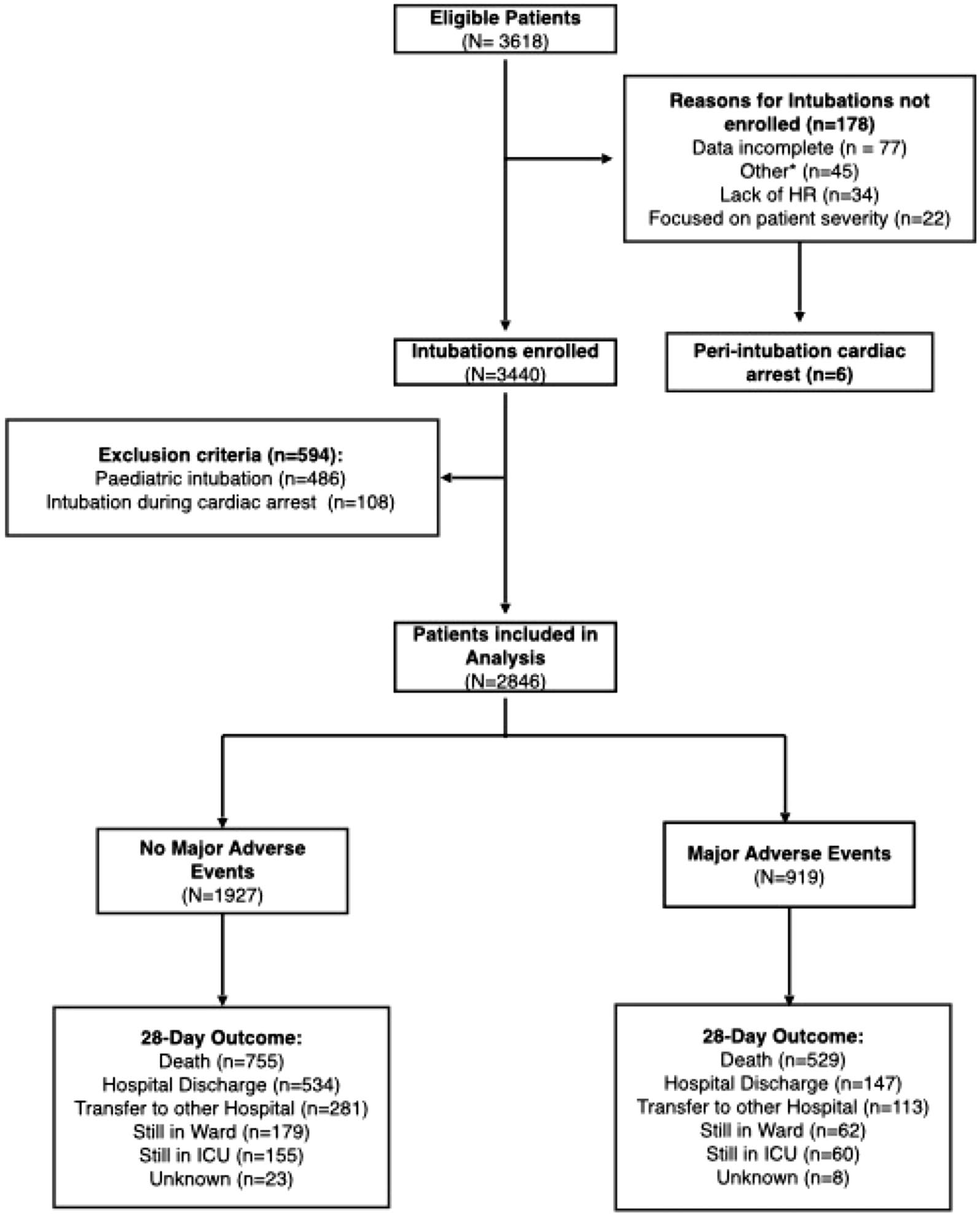
Fig. 1 Enrollment flow diagram. *Others reasons for no inclusion intubations were “forgot to fill out”, “doesn’t remember completing”, or “invalid record number”). HR: Health Records; ICU: Intensive Care Unit
Maia et al. Critical Care (2025) 29:155 Page 5 of 15
Patient baseline characteristics
The median age was 63 years (IQR, 49–73), and most were male (58.0%). Patients who experienced MAEs were older (median age 65 vs 62 years; p < 0.001), had a higher pre-intubation shock index (0.87 vs 0.74; p < 0.001), and were more likely to have been intubated for acute respira-tory failure (47.9% vs 30.6%, p < 0.001). Table 1 and eTa-ble 2 detail the characteristics of included patients.
Intubation characteristics
First-attempt success occurred in 2116 (74.3%) cases. Direct laryngoscopy (DL) was used for the first intuba-tion attempt in 2294 (80.6%) cases (Table 1). The median number of attempts was 1 (IQR, 1–2), with difficult intu-bations occurring in 198 (7.0%) patients. Pre-treatment with fentanyl was used in 630 (22.1%) intubations. Eto-midate was the most used induction agent (n= 1655 [58.2%]), followed by ketamine (n= 751 [26.4%]). Propo-fol was rarely used (n= 89 [3.1%]). Rapid sequence intubation (RSI) was employed in 2462 (86.5%) of the intubations. Residents in Emergency Medicine and Inter-nal Medicine performed the first intubation attempt in 1097 (38.5%) and 1125 (39.5%) cases, respectively. As first operators, anesthesiologists participated in only 3 intuba-tions (0.1%). First-year residents were the first operators in 1351 (47.5%) cases. A surgical airway was performed on only 1 (0.04%).
Incidence of MAEs
Of 2846 patients, 919 (32.3%) experienced at least one MAEs. New hemodynamic instability was the most com-mon MAE (569, 20.0%), followed by severe hypoxemia in 356 (12.5%) intubations, and peri-intubation cardiac arrest in 100 (3.5%) intubations, with 73 (73.0%) of these achieving the return of spontaneous circulation. Table 2 presents other complications. At least one episode of transient hypotension occurred in 345 (12.1%) intuba-tions, and esophageal intubation was reported in 86 (3.0%) intubations.
Association of MAEs with 28‑day mortality
Overall, 28-day mortality was 45.1%. Mortality was higher among patients experiencing MAEs compared to those who did not (57.6 vs 39.2%, p < 0.001). Figures 2 and 3 presents the association of MAEs and their sub-components with 28-day mortality. MAEs were associ-ated with increased 28-day mortality (aHR 1.43, 95% CI 1.26–1.62, p < 0.001). All subcomponents were also associated with increased 28-day mortality, including increased HR from hemodynamic instability (aHR 1.28, 95% CI 1.11–1.48, p < 0.001) followed by severe hypox-emia (adjHR 1.39, 95% CI, 1.16–1.66, p < 0.001) and cardiac arrest (aHR 2.52, 95% CI 1.86–3.40, p < 0.001).
Finally, patients experiencing both hemodynamic insta-bility and severe hypoxemia had an increased risk of mortality compared to those with neither (aHR 1.97, 95% CI 1.38–2.81, p < 0.001, Appendix file, eTable 7), which was higher than either hemodynamic instability or severe hypoxemia alone.
All sensitivity analyses to the Cox model assumptions demonstrated similar results (Fig. 2 and Appendix, eTa-bles 8–11), suggesting the robustness of model assump-tions. E-values for the point estimate and its lower confidence interval were, respectively, 1.88 and 1.63 for all MAEs; 1.66 and 1.36 for hemodynamic instability; 1.82 and 1.45 for severe hypoxemia; and 3.18 and 2.44 for cardiac arrests (Appendix file, eFigures 4–7).
Factors associated with MAEs and first‑attempt success
Patients who experienced MAEs had lower observed first-attempt success (66.3% vs 78.2%, p < 0.001). Success-ful first-attempt intubation was associated with a lower likelihood of experiencing MAEs (aOR 0.52, 95% CI 0.41–0.65, p < 0.001), while each additional attempt was associated with higher odds of MAEs (aOR 1.65, 95% CI: 1.45–1.88, p < 0.001; Appendix file, eTable 5). Moreover, each additional intubation attempt markedly increased the risk of severe hypoxemia (aOR 2.28, 95% CI 1.95–2.65, p < 0.001, eTable 5). Increasing age, elevated shock index, higher SOFA score, lower pre-intubation oxygen saturation, and acute respiratory failure as an indication for intubation were significantly associated with peri-intubation MAEs in the multivariable analysis (Table 3). Performing an intubation checklist was significantly pro-tective against peri-intubation MAEs (aOR, 0.75; 95% CI 0.57–0.98; p= 0.036). Patients receiving ketamine expe-rienced a higher unadjusted rate of MAEs compared to those receiving etomidate (29.8 vs 24.8%, p < 0.001), however, no significant difference in MAEs was observed among different induction agents in multivariable analy-sis (Table 3). The specialty of the first operator was asso-ciated with first-attempt success, with internal medicine (OR 0.61, 95% CI 0.44–0.83, p= 0.002) specialists having lower odds of first-attempt success compared to emer-gency medicine specialists. Intubation with a bougie was associated with higher odds of first-attempt success (OR 1.57, 95% CI 1.06–2.34, p= 0.025, eTable 6).
Discussion
In this multicenter prospective cohort study of criti-cally ill adults undergoing emergency tracheal intuba-tion, MAEs occurred in 32.3% of patients: hemodynamic instability, in 20.0%; severe hypoxemia, in 12.5%; and cardiac arrest, in 3.5%. The overall 28-day mortality was 45.1%. Patients who experienced MAEs had significantly higher mortality, even after adjustment for patient-level
Maia et al. Critical Care (2025) 29:155 Page 6 of 15
Table 1 Patient demographics and intubation characteristics
All patients Major Adverse event No major events p-value
(N = 2846) (N = 919) (N = 1927)
Patient Characteristics
Age, years, median (IQR) 63 (49, 73) 65 (51, 75) 62 (47, 72) < 0.001
Sex, male, n (%) 1652 (58%) 543 (59.1%) 1109 (57.6%) 0.44
BMI, kg/m2, median (IQR) 25.7 (23.4, 27.8) 25.7 (23.4, 27.8) 25.7 (23.4, 27.8) 0.74
Data available, n (%) 2823 (99.2%) 912 (99.2%) 1911 (99.2%)
Mean arterial pressure, mmHg, median (IQR) 96.7 (80, 113.3) 89.8 (76, 106.7) 99.3 (83.3, 116.7) < 0.0001
Data available, n (%) 2764 (97.1%) 876 (95.3%) 1888 (98%)
Shock index, median (IQR) 0.78 (0.61, 0.99) 0.87 (0.68, 1.08) 0.74 (0.58, 0.93) < 0.0001
Data available, n (%) 2756 (96.8%) 873 (95%) 1883 (97.7%)
Charlson comorbidity index, 0–37, median (IQR) 3 (1, 5) 3 (2, 5) 3 (1, 5) < 0.0001
SOFA score*, 0–24, median (IQR) 4 (3, 6) 4 (3, 7) 4 (2, eh) < 0.0001
Hemodynamic resuscitation preintubation, n (%)
Fluidsa 616 (21.6%) 246 (26.8%) 370 (19.2%) < 0.0001
Vasopressorsb 831 (29.2%) 331 (36%) 500 (25.9%) < 0.0001
Blood transfusion 65 (2.3%) 31 (3.4%) 34 (1.8%)
Intubation Characteristics
Indication for intubation, n (%) < 0.0001
Airway protection 1292 (45.4%) 310 (33.7%) 982 (51.0%)
Acute respiratory failure 1029 (36.2%) 440 (47.9%) 589 (30.6%)
Anticipation of clinical 449 (15.8%) 151 (16.4%) 298 (15.5%)
course
Transport risk 13 (0.5%) 1 (0.1%) 12 (0.6%)
Not recorded 63 (2.2%) 17 (1.8%) 46 (2.4%)
Primary diagnosis of trauma, n (%) 222 (7.8%) 55 (6.0%) 167 (8.7%) 0.013
Subjective impression of difficult intubationc, n (%) 762 (26.8%) 287 (31.2%) 475 (24.6%) < 0.0001
Time between indication and intubation**, n (%) 0.026
0–15 min 1430 (50.2%) 424 (46.1%) 1006 (52.2%)
15–60 min 1189 (41.8%) 414 (45%) 775 (40.2%)
Unknown timing 5 (0.2%) 2 (0.2%) 3 (0.2%)
Institutional checklist performed prior to intubation, n (%) 0.011
No 1593 (56.0%) 546 (59.4%) 1047 (54.3%)
Yes 1253 (44%) 373 (40.6%) 880 (45.7%)
Preoxygenation method, n (%)
Non-invasive ventilation 353 (12.4%) 134 (14.6%) 219 (11.4%)
Non-rebreathing mask 870 (30.6%) 252 (27.4%) 618 (32.1%)
Bag-Valve-Mask 1514 (53.2%) 498 (54.2%) 1016 (52.7%)
High-flow nasal catheter 47 (1.7%) 10 (1.1%) 37 (1.9%)
Other preoxygenation 35 (1.2%) 12 (1.3%) 23 (1.2%)
No preoxygenation 26 (0.9%) 13 (1.4%) 13 (0.7%)
Not recorded 1 (0%) 0 (0%) 1 (0.1%)
SpO2 measured before intubation, % 99 (95, 100) 97 (92, 100) 99 (96, 100) < 0.0001
Intubation method, n (%) 0.28
RSI 2389 (83.9%) 758 (82.5%) 1631 (84.6%)
DSI 183 (6.4%) 70 (7.6%) 113 (5.9%)
No paralytics 225 (7.9%) 73 (7.9%) 152 (7.9%)
Other methodd 47 (1.7%) 18 (2%) 29 (1.5%)
Unknown/Not recorded 2 (0.1%) 0 (0%) 2 (0.1%)
First attempt device, n (%) 0.46
Maia et al. Critical Care (2025) 29:155 Page 7 of 15
Table 1 (continued)
All patients Major Adverse event No major events p-value
(N = 2846) (N = 919) (N = 1927)
DL—Curved 2294 (80.6%) 733 (79.8%) 1561 (81.0%)
VL—Standard 444 (15.6%) 154 (16.8%) 290 (15.0%)
VL—Hyperangulated 73 (2.6%) 25 (2.7%) 48 (2.5%)
Other devicee 29 (1%) 6 (0.7%) 23 (1.2%)
Unknown 6 (0.2%) 1 (0.1%) 5 (0.3%)
Auxiliary intubation device, n (%) 0.14
Stylet 1466 (51.5%) 450 (49.0%) 1016 (52.7%)
Bougie 923 (32.4%) 316 (34.4%) 607 (31.5%)
None 443 (15.6%) 151 (16.4%) 292 (15.2%)
Other 8 (0.3%) 2 (0.2%) 6 (0.3%)
Unknown/Not recorded 6 (0.2%) 0 (0%) 6 (0.3%)
Capnography for intubation confirmation, n (%) 637 (22.4%) 196 (21.3%) 441 (22.9%) 0.51
Cormack-Lehane classification, n (%) 0.003
1 1281 (45%) 380 (41.3%) 901 (46.8%)
2a 830 (29.2%) 267 (29.1%) 563 (29.2%)
2b 443 (15.6%) 160 (17.4%) 283 (14.7%)
3 162 (5.7%) 62 (6.7%) 100 (5.2%)
4 17 (0.6%) 11 (1.2%) 6 (0.3%)
Not available 113 (4.0%) 39 (4.2%) 74 (3.8%)
Apneic oxygenation performed, n (%) 0.009
No 2025 (71.2%) 678 (73.8%) 1347 (69.9%)
Yes 628 (22.1%) 197 (21.4%) 431 (22.4%)
Unknown 193 (6.8%) 44 (4.8%) 149 (7.7%)
Induction analgesia, n (%) 0.014
Fentanyl 630 (22.1%) 172 (18.7%) 458 (23.8%)
Lidocaine 42 (1.5%) 19 (2.1%) 23 (1.2%)
Other 99 (3.5%) 31 (3.4%) 68 (3.5%)
None 2074 (72.9%) 697 (75.8%) 1377 (71.5%)
Unknown 1 (0%) 0 (0%) 1 (0.1%)
Induction hypnotic, n (%) < 0.0001
Etomidate 1655 (58.2%) 499 (54.3%) 1156 (60.0%)
Ketamine 751 (26.4%) 274 (29.8%) 477 (24.8%)
Midazolam 145 (5.1%) 32 (3.5%) 113 (5.9%)
Propofol 89 (3.1%) 29 (3.2%) 60 (3.1%)
Otherf 16 (0.6%) 7 (0.8%) 9 (0.5%)
None 91 (3.2%) 44 (4.8%) 47 (2.4%)
Unknown 99 (3.5%) 34 (3.7%) 65 (3.4%)
Induction neuromuscular blocker, n (%) 0.11
Succinylcholine 1589 (55.8%) 494 (53.8%) 1095 (56.8%)
Rocuronium 1021 (35.9%) 353 (38.4%) 668 (34.7%)
None 219 (7.7%) 70 (7.6%) 149 (7.7%)
Otherg 14 (0.5%) 1 (0.1%) 13 (0.7%)
Unknown 3 (0.1%) 1 (0.1%) 2 (0.1%)
First operator specialty, n (%) 0.013
Internal Medicine 1125 (39.5%) 407 (44.3%) 718 (37.3%)
Emergency Medicine 1097 (38.5%) 323 (35.1%) 774 (40.2%)
Surgery 156 (5.5%) 47 (5.1%) 109 (5.7%)
Medical student 102 (3.6%) 22 (2.4%) 80 (4.2%)
ICU 42 (1.5%) 13 (1.4%) 29 (1.5%)
Maia et al. Critical Care (2025) 29:155 Page 8 of 15
Table 1 (continued)
All patients Major Adverse event No major events p-value
(N = 2846) (N = 919) (N = 1927)
Anesthesiology 3 (0.1%) 1 (0.1%) 2 (0.1%)
Other specialtyh 177 (6.2%) 60 (6.5%) 117 (6.1%)
Unknown/Not recorded 144 (5.1%) 46 (5.0%) 98 (5.1%)
First operator training stage, n (%) < 0.0001
Resident, 1 st year 1351 (47.5%) 430 (46.8%) 921 (47.8%)
Resident, 2nd year 719 (25.3%) 211 (23.0%) 508 (26.4%)
Resident, 3rd year 112 (3.9%) 45 (4.9%) 67 (3.5%)
Resident, 4 th year 2 (0.1%) 1 (0.1%) 1 (0.1%)
Attending physician 409 (14.4%) 131 (14.3%) 278 (14.4%)
Medical student 120 (4.2%) 29 (3.2%) 91 (4.7%)
Other operatori 11 (0.4%) 3 (0.3%) 8 (0.4%)
Unknown/Not recorded 122 (4.3%) 69 (7.5%) 53 (2.8%)
First-attempt intubation success, n (%) 2116 (74.3%) 609 (66.3%) 1507 (78.2%) < 0.0001
Difficult intubation, n (%) 198 (7%) 110 (12%) 88 (4.6%) < 0.0001
IQR: Interquartile range; SD: Standard deviations; RSI: Rapid sequence intubation; DSI: Delayed sequence intubation; BMI: Body mass index: calculated as weight in
kilograms divided by height in meters squared; ICU: Intensive care unit; DL: Direct laryngoscopy; VL: Video laryngoscopy; TI: Tracheal intubation; SOFA: Sequential
organ failure assessment
for SpO2 < 92%. Scores of 3 and 4 were not applicable as no patients were on mechanical ventilation
a: Administration of a fluid bolus > 15 mL/kg to maintain target blood pressure
b: New requirement for or at least 25% increase in the dose of vasopressor
c: Subjective, global clinical assessment made by the operator before the intubation
d: Alternative methods of intubation, such as awake intubation and nasal intubation
e: Other devices such as fiberoptic intubation, Airway Scope, Airtraq
f: Other possible agents are thiopental and dexmedetomidine
g: Atracurium, cisatracurium, pancuronium and vecuronium are other options
h: Included are all other medical specialties that do not match the above categories, such as neurologists, psychiatrists, and others
i: Included career medical officers, senior medical officers and paramedics
and intubation-related characteristics. These associations remained consistent across multiple sensitivity analyses. Factors associated with MAEs included both nonmodifi-able characteristics and potentially modifiable elements, such as first-attempt success, pre-intubation hemo-dynamic status, and pre-intubation peripheral oxygen saturation.
In 2021, INTUBE study analyzed 2964 intubations in critically ill patients and observed MAEs in 45.2% of cases, a higher incidence compared to 32.3% in our study. They reported new hemodynamic instability in 42.6% of patients, severe hypoxemia in 9.3%, and cardiac arrest during intubation in 3.1% [4]. In contrast, our cohort experienced less new hemodynamic instability (20.0%), and more severe hypoxemia (12.3%) and cardiac arrest (3.5%). In a different population, with slight differences in the definition of MAE, the PREPARE II trial reported findings that were similar to our observations, with an incidence of cardiovascular collapse at 21.0% [20]. In our cohort, the utilization of propofol as an induction agent
(3%) and opioids (18%) prior to intubation was nota-bly lower compared to INTUBE cohort (45% and 51%, respectively) [4]. Propofol, while widely used for its rapid onset and short duration of action, is associated with a higher incidence of hemodynamic instability [21, 22]. In a secondary analysis from INTUBE study, propofol was identified as a modifiable factor linked to increased peri-intubation hemodynamic instability, with an aOR of 1.28 [2]. Furthermore, the practice of using opioids as a pre-treatment during emergency intubation is con-troversial. North American registry cohorts with 15,776 intubations show that opioids were used in only 2.9% of patients [6]. Despite limitations, Ferguson I et al. raised questions about the risk of hypotension when fentanyl is used as pre-treatment during emergency intubation [23]. Differences in drug preferences likely reflect global varia-tions in clinical practice, as illustrated by the significantly higher proportion of anesthesiologists performing intu-bations in INTUBE cohort (54%) compared to our study (0.4%) [4]. Although the Brazilian model of outsourcing
Maia et al. Critical Care (2025) 29:155 Page 9 of 15
Table 2 Frequency of peri-intubation adverse events
All patients
(N = 2846)
Any major adverse event 919 (32.3%)
New hemodynamic instability 569 (20.0%)
Type of hemodynamic instability (n = 569)
Systolic BP ≤ 65 mmHg 340 (59.8%)
Needed new vasopressor 272 (47.8%)
Increased dose of vasopressor 183 (32.2%)
Push dose of vasopressor 32 (5.6%)
Needed fluid therapy ≥ 15 ml/kg 34 (6.0%)
Severe hypoxemia 356 (12.5%)
Cardiac arrest 100 (3.5%)
Cardiac arrest immediate outcome (n = 100)
Death 26 (26.0%)
Return of spontaneous circulation 73 (73.0%)
Unknown 1 (1.0%)
Other adverse events
Transient hypotension 345 (12.1%)
Esophageal intubation 81 (2.8%)
(immediate identification, < 5 min)
Arrhythmia 50 (1.8%)
Aspiration 25 (0.9%)
Tooth trauma 12 (0.4%)
Airway injury 7 (0.2%)
Esophageal intubation 5 (0.2%)
(delayed identification, > 5 min)
Pneumomediastinum 2 (0.1%)
Vocal cord avulsion 1 (0.0%)
BP: Blood pressure
anesthesia services differs from that in Europe, in many North American centers, anesthesiologists do not com-monly perform intubations outside the operating room either [14]. The generalization of this finding depends on the contextual factors of each country regarding airway management operators and specialties.
In our sample, patients experiencing MAEs had a mark-edly higher 28-day mortality, with an aHR of 1.43 (95% CI 1.26–1.62). These findings align with those reported by Russoto et al., who also found an association between MAEs and 28-day mortality (aOR 1.44, 95% CI 1.19–1.74) [4]. While recent trials have addressed modifiable factors to improve pre-oxygenation and avoid hypoxemia [12] or enhance first-attempt success [14, 24], factors to prevent cardiovascular collapse have not been extensively investigated or were not effective [20, 25]. In our cohort, patients who received fluid resuscitation or vasopres-sors prior to intubation were more likely to experience MAEs, suggesting a more severe clinical profile at base-line. Although these findings highlight the importance of hemodynamic optimization before intubation, it is also
possible that hemodynamic instability serves as a surro-gate marker of illness severity not fully captured by the available covariates. This interpretation is supported by the lower E-values observed for hemodynamic instability compared with other MAEs, indicating greater suscepti-bility to unmeasured confounding.
Recent consensus guidelines from an international Del-phi study highlight structured strategies for managing the physiologically difficult airway, emphasizing pre-intuba-tion assessment, hemodynamic stabilization, optimized pre-oxygenation, and careful induction agent selection to minimize peri-intubation adverse events [8]. We identi-fied several key risk factors for MAEs during intubation, consistent with previous research. De Jong et al. high-lighted hypotension, hypoxemia, lack of pre-oxygena-tion, obesity, and age over 75 years as critical risk factors for cardiac arrest [26]. In our cohort, markers of illness severity, including shock index, the number of organ dys-functions, and vasopressor use, were strongly associated with MAEs. Therefore, targeted efforts to identify and stabilize patients presenting with shock or respiratory failure before intubation are essential. Early optimiza-tion of hemodynamic status and oxygenation may reduce the risk of peri-intubation adverse events. Two ongo-ing international trials, the FLUVA (NCT05318066) and PREVENTION (NCT05014581) trials, are evaluating if pre-emptive vasopressor use can reduce cardiovascular collapse during intubation in critically ill adults and may help clarify the role of hemodynamic optimization in this context. Furthermore, capnography use was limited in our cohort (25%), similar to INTUBE study4, and likely reflects restricted access to waveform capnography in many resource-limited settings. In contrast, use of a pre-intubation checklist was associated with a lower risk of MAEs (aOR, 0.75), supporting its value as a simple, high-impact intervention to improve the safety of emergency airway management.
Our findings highlight the protective effect of first-attempt intubation success in reducing MAEs (aOR, 0.52), with each additional intubation attempt signifi-cantly increasing the odds of severe hypoxemia (aOR, 2.28). Operator specialty was significantly associated with first-attempt success, emergency medicine resi-dents achieved higher success rates, emphasizing the importance of specialized training in emergency air-way management. Although anesthesiologists are con-sidered airway, they performed very few intubations in our cohort (3 first-attempt intubations and 11 attempts overall), limiting meaningful comparisons with other specialties. Despite evidence supporting the use of vide-olaryngoscopes and bougies to improve first-attempt success [27, 28] these devices were underutilized in our cohort, similar to the INTUBE4 cohort in which
Maia et al. Critical Care (2025) 29:155 Page 10 of 15
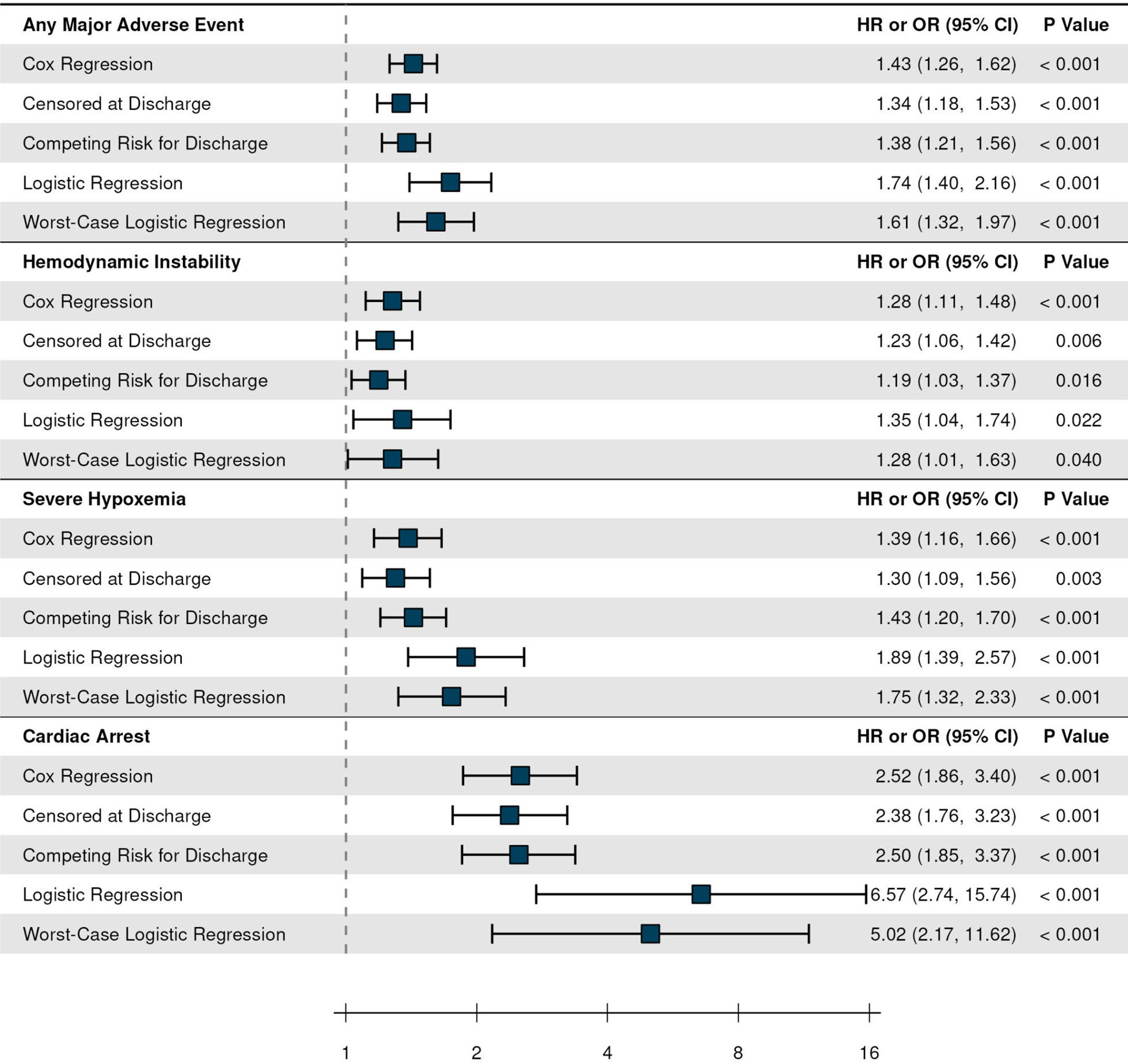
Fig. 2 Association between the occurrence of major adverse events and 28-day mortality. HR: Hazard Ratio; OR: Odds Ratio; CI: Confidence Interval; MAE: Major Adverse Event; Major adverse events peri-intubation were defined as events during or thirty minutes after the intubation process, as listed below: hemodynamic Instability: Systolic Blood Pressure < 65 mmHg, need for starting or increase in vasopressor dose or fluid resuscitation; Severe hypoxemia: Oxygen peripheral saturation less than 80%; Cardiac arrest: Presence of cardiac arrest signs peri-intubation
videolaryngoscopy was used in only 17% of cases. This limited use, along with the high proportion of novice first operators, may have contributed to the lower first-attempt success rate observed in our study (74.3%). The higher incidence of severe hypoxemia (12.5%) compared with Russotto et al4 may also be explained by this lower first-attempt success rate, as well as the predominant use of bag-valve-mask ventilation for preoxygenation, rather than noninvasive ventilation or high-flow nasal oxygen. Although evidence suggests that stylet use may reduce
complications and improve first-attempt success, 15% of patients in our cohort were intubated without any auxil-iary devices [29]. Future efforts should focus on improv-ing access to these devices in LMICs.
The choice of induction agent for intubation remains an important area of investigation. In this study, eto-midate was associated with fewer MAEs in univariate analysis; however, the association was not significant in the multivariable analysis. The survival benefit of etomi-date remains uncertain, and recent evidence, including
Maia et al. Critical Care (2025) 29:155 Page 11 of 15
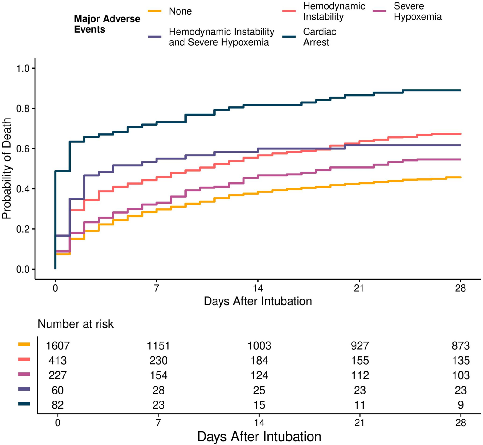
Fig. 3 Mortality rate by days after intubations, stratified by major adverse events. Patients who were discharged were considered alive
through the 28-day follow-up period for this analysis
a meta-analysis by Kotani et al., has reported findings favoring ketamine [30–33]. Further research is needed to clarify the comparative effectiveness of induction agents in critically ill patients..
Strengths and limitations
Our study has several strengths. As the first prospective airway cohort established in a low- and middle-income country, this research captures variability in practice beyond what is typically observed in high-income coun-tries. Additionally, we adhered to current best practices in causal inference, carefully selecting confounders and conducting sensitivity analyses to address both mod-eling assumptions and unmeasured confounding, which enhances the interpretability of our findings.
However, our study has limitations. First, we did not follow up with patients after hospital discharge, which may result in an underestimation of mortality. To address this, we conducted multiple sensitivity analy-ses to address potential informative censoring, and the
effect estimates remained consistent. We did not assess longer-term outcomes either, which would be a gap for future research. Second, despite adjusting for multiple confounders for the association of MAEs with 28-day mortality, the possibility of residual confounding can-not be entirely excluded, particularly in the context of hemodynamic instability, which was more susceptible to unmeasured confounding. Importantly, the other observed associations of multivariable analyses should be interpreted as exploratory to guide future research. Third, although each center had a case manager, not all consecutive intubations were captured in the partici-pating centers. Nonetheless, the incidence of cardiac arrest among non-enrolled patients was comparable to that among enrolled patients, suggesting minimal selection bias. Fourth, while the center enrollment process was broad, most participating centers were academic institutions with emergency residency pro-grams, which may have led to a more selected sample with potentially higher standards of care compared to
Maia et al. Critical Care (2025) 29:155 Page 12 of 15
Table 3 Association between patient and intubation characteristics with peri-intubation major adverse events
Univariable Multivariable
Characteristic Odds ratio p value Odds ratio1 p value
(95% CI) (95% CI)
Age, per 5 years 1.04 (1.02, 1.07) 0.001 1.05 (1.01, 1.09) 0.019
Sex, male 1.08 (0.92, 1.28) 0.33 –
BMI, per 5 kg/m2 1.00 (0.98, 1.01) 0.59 –
Shock index, per 0.1 point 1.13 (1.10, 1.16) < 0.001 1.10 (1.06, 1.13) < 0.001
Charlson comorbidity, per 1 point 1.07 (1.03, 1.10) < 0.001 1.05 (0.99, 1.10) 0.091
SOFA score, per 1 point 1.08 (1.05, 1.11) < 0.001 1.07 (1.03, 1.11) < 0.001
Indication for intubation
Airway protection -Reference- –- -Reference-–-
Acute respiratory 2.41 (2.01, 2.89) < 0.001 1.65 (1.27, 2.14) < 0.001
insufficiency
Anticipation of 1.66 (1.31, 2.11) < 0.001 1.27 (0.91, 1.75) 0.16
clinical course
Transport risk 0.26 (0.03, 2.06) 0.20 –
MACOCHA score, per 1 point 0.95 (0.88, 1.03) 0.20–
MACOCHA criteria
Mouth opening < 3 cm 0.84 (0.51, 1.36) 0.47–
Cervical restriction 1.48 (1.14, 1.92) 0.003 1.18 (0.82, 1.68) 0.37
Obstructive apnea 1.32 (0.81, 2.12) 0.26–
Glasgow score < 9 0.72 (0.60, 0.87) < 0.001 1.09 (0.84, 1.43) 0.51
Mallampati grade 3 or 4 0.98 (0.40, 2.40) 0.96–
Subjective impression of difficult airway 1.34 (1.12, 1.60) 0.001 1.22 (0.95, 1.57) 0.13
Pre-intubation oxygen saturation, per % 0.94 (0.93, 0.95) < 0.001 0.94 (0.93, 0.94) < 0.001
Intubation checklist performed 0.74 (0.60, 0.91) 0.005 0.75 (0.57, 0.98) 0.036
Intubation method
RSI -Reference- –-Reference–-
DSI 1.21 (0.88, 1.66) 0.23 –
Other oral intubation 1.26 (0.86, 1.85) 0.23 –
Non-oral intubation 1.82 (0.94, 3.54) 0.077 –
First-attempt—visualization device
DL—Curved -Reference- – – –
VL—Standard or 1.24 (0.99, 1.55) 0.064 –
Hyperangulated
Other device 0.62 (0.25, 1.55) 0.31 –
Auxiliary intubation device
None -Reference- – Reference –
Bougie 1.07 (0.82, 1.39) 0.63 1.22 (0.86, 1.73) 0.26
Stylet 0.75 (0.58, 0.96) 0.021 0.95 (0.68, 1.33) 0.76
Other 0.55 (0.11, 2.85) 0.48 0.34 (0.04, 3.37) 0.36
Apneic oxygenation performed 1.00 (0.81, 1.23) 0.98 –
Cormack-Lehane Class
1-Reference- – -Reference- –
2a 1.12 (0.92, 1.35) 0.26 0.94 (0.74, 1.20) 0.62
2b 1.39 (1.10, 1.76) 0.005 1.03 (0.76, 1.41) 0.84
3 1.55 (1.10, 2.19) 0.013 1.00 (0.62, 1.61) 0.98
4 4.61 (1.67, 12.8) 0.003 2.38 (0.61, 1.05) 0.21
Induction analgesia
None -Reference- – -Reference- –
Any analgesia 0.80 (0.66, 0.97) 0.024 0.81 (0.62, 1.05) 0.11
Maia et al. Critical Care (2025) 29:155 Page 13 of 15
Table 3 (continued)
Univariable Multivariable
Characteristic Odds ratio p value Odds ratio1 p value
(95% CI) (95% CI)
Induction hypnotic
Etomidate -Reference- – Reference–
Ketamine 1.44 (1.18, 1.75) < 0.001 1.24 (0.94, 1.62) 0.13
Midazolam 0.77 (0.49, 1.19) 0.24 1.49 (0.73, 3.05) 0.27
Propofol 1.13 (0.71, 1.80) 0.60 1.61 (0.85, 3.04) 0.15
Other hypnotic 1.78 (0.64, 4.89) 0.27 1.35 (0.39, 4.75) 0.64
None 2.37 (1.53, 3.65) < 0.001 1.07 (0.70, 1.61) 0.76
Induction paralytic use
None -Reference- – Reference–
Any paralytic 0.75 (0.52, 1.07) 0.11 –
First operator specialty
Emergency Medicine -Reference- –- Reference–-
Internal Medicine 1.14 (0.92, 1.42) 0.23 –
Surgery 0.86 (0.58, 1.28) 0.46–
Medical Student 0.65 (0.39, 1.08) 0.098–
ICU 0.96 (0.49, 1.89) 0.91–
Other specialty 1.16 (0.81, 1.66) 0.43–
First operator age, per year 1.00 (0.98, 1.01) 0.66–
First operator experience, per year 1.00 (0.98, 1.02) 0.91–
First operator—time on call, per 5 h 1.01 (0.94, 1.09) 0.81–
1 Multivariable model included a LASSO penalty for variable selection. Odds ratios for characteristics excluded from the final model are listed as “–"
Abbreviations: BMI: Body mass index: calculated as weight in kilograms divided by height in meters squared; SOFA: Sequential organ failure assessment; ICU: Intensive
care unit; RSI: Rapid sequence intubation; DSI: Delayed sequence intubation; DL: Direct laryngoscopy; VL: Videolaryngoscopy
other centers in Brazil. Fifth, we did not collect specific data on withdrawal of care, which limits our ability to evaluate its impact on 28-day mortality, although there is no strong reason to assume that withdrawal decisions were directly influenced by peri-intubation adverse events, except in cases of peri-intubation cardiac arrest. Furthermore, we did not record the annual rate of intu-bations performed at each site, limiting our ability to assess how intubation volume or operator experience could have influenced outcomes. Moreover, the inclu-sion of patients exclusively from emergency depart-ments may be a limitation, as the findings may not fully reflect outcomes among all critically ill patients, par-ticularly regarding the incidence of adverse events and 28-day mortality. However, in our setting, critically ill patients are often intubated in the ED before an ICU bed becomes available. As such, these results provide important insights into the quality of pre-ICU care and highlight opportunities for improvement during this critical period. Lastly, a few centers were rural units without ICU beds, so patients were transferred after stabilization, and we did not have access to 28-day hos-pital outcomes for these patients.
Conclusions
In conclusion, one in three patients experienced a peri-intubation major adverse event, which may increase 28-day mortality. First-attempt success, pre-intubation hemodynamics, pre-oxygenation, and sedative choices are potentially modifiable factors that may reduce the risk of MAEs. These findings highlight an urgent need for targeted interventions to mitigate peri-intubation adverse events in resource-constrained settings.
Ethics approval and consent to participate
This study was approved by the ethics committees of all participating centres. Due to the observational nature of the study and the use of de-identified data, a waiver of individual patient consent was granted by each institution’s ethics committee. Certificate of Presenta-tion for Ethical Consideration at Coordinator Center: CAE-52424821.1.0000.0068.
Maia et al. Critical Care (2025) 29:155 Page 14 of 15
Consent for publication
Not applicable.
Supplementary Information
The online version contains supplementary material available at .
Additional file1
Acknowledgements
BARCO Group: Victor Paro da Cunha, MD; Julio F. Marchini, MD, PhD; Patricia Albuquerque Moura, RT; Fernanda Greco, RT; Yasmine Filippo, RT; Rubens Yoshinori Kai, MD; Guilherme Torres Abi Ramia Chimelli, MD; Juan Valdivia, MD; Edson Luiz Favero Junior, MD, PhD; Felipe Rischini, MD; Vitor Câmara, MD; Hen-rique Bertotto, MD; Victor Borges, MD; Juliano Rathke, MD; Renato Melo, MD; Ariadine Augusta Maiante, MD; Sarah Maciel Silva, MD; Clarisse Moreira Ribeiro de Oliveira, MD; Andressa Pi Rocha Reis, MD; Thamyres de Carvalho Rufato, MD; Gabriella Dias, MD; Victoria Sartor Poloni, MD; Kauê Lima, MD; Hilana Zenly, MD; José Carlos Motta, MD; Gabriel Miranda, MD; Alexandre Freitas, MD; Leonardo Gasperini, RT; Thais Raimondi Sudbrack, MD; Ana Paula Ribeiro, MD; Eduardo Mensch Jaeger, MD; Guilherme Henrique A. do Carmo, MD; Andrea de Vargas Tomelero, MD; Augusto Lengler Konrath, MD; Vitor Cremonese Zanella, MD; Natalia Fuhr, MD; Davi Amaral Cesário Rosa, MD; Isabela Lopes Lima, MD; Luiz Fernando Varela, MD; Isabella Baldino, MD; Andre Zimmerman, MD, PhD; Julia M. Dorn de Carvalho, MD; Molly M. Jeffrey, PhD.
Author contributions
Ian Ward A. Maia, Gabriela Stanzani, Heraldo P. Souza, and Julio C. G. Alencar conceived and designed the study. All authors participated in data collection.
Ian Ward A. Maia and Aidan Mullan performed the data analysis. Ian Ward A. Maia drafted the manuscript, and all authors contributed to the draft. Ian Ward A. Maia, Otavio T. Ranzani, Fernanda Bellolio, Lucas Oliveira J. e Silva, Rafael von Hellmann, Bruno A. M. Pinheiro Besen, and Julio C. G. Alencar critically reviewed and revised the manuscript. All authors approved the final version and are accountable for all aspects of the work.
Rochester, MN, USA. 3 Medical Sciences Postgraduate Program, Faculdade de Medicina, Universidade de Sao Paulo, Sao Paulo, Brazil. 4 Department of Emer-gency Medicine, Universidade Federal do Rio Grande do Sul, Hospital de Clini-cas de Porto Alegre, Porto Alegre, Rio Grande Do Sul, Brazil. 5 Intensive Care Department, Monash Health, Melbourne, Australia. 6 Department of Emer-gency Medicine, Hospital Metropolitano Odilon Behrens, Belo Horizonte, Minas Gerais, Brazil. 7 Department of Emergency Medicine, Ribeirao Preto School of Medicine-University of Sao Paulo, Ribeirao Preto, São Paulo, Brazil. 8 Department of Emergency Medicine, Hospital das Clínicas da Faculdade de Medicina de Botucatu, Botucatu, Sao Paulo, Brazil. 9 Department of Emergency Medicine, Hospital Regional de Sao José - Dr. Homero de Miranda Gomes, São José, Santa Catarina, Brazil. 10 Department of Emergency Medicine, Hospital Sao Lucas da Pontificia Universidade Católica Do Rio Grande Do Sul, Porto Alegre, Rio Grande Do Sul, Brazil. 11 Department of Emergency Medicine, Hospital de Pronto Socorro, Porto Alegre, Rio Grande Do Sul, Brazil. 12 Depart-ment of Emergency Medicine, Hospital Geral de Fortaleza, Fortaleza, Ceara, Brazil. 13 Department of Emergency Medicine, Hospital Dr. Carlos Alberto Studart Gomes, Fortaleza, Ceara, Brazil. 14 Department of Emergency Medicine, Hospital das Clínicas da Faculdade de Medicina de Marilia, Marilia, Sao Paulo, Brazil. 15 Department of Emergency Medicine, Hospital Augusto de Oliveira Camargo, Indaiatuba, Sao Paulo, Brazil. 16 Department of Emergency Medicine, Hospital Regional Alto Vale, Rio Do Sul, Santa Catarina, Brazil. 17 Department of Emergency Medicine, Hospital Santo Antonio, Sinop, Mato Grosso, Brazil. 18 Department of Emergency Medicine, Hospital Nossa Senhora da Conceição, Porto Alegre, Rio Grande Do Sul, Brazil. 19 Department of Emergency Medicine, Hospital Bruno Born, Lajeado, Rio Grande Do Sul, Brazil. 20 Unidade de Pronto Atendimento de Lajeado, Lajeado, Rio Grande Do Sul, Brazil. 21 Department of Emergency Medicine, Hospital Santa Cruz, Santa Cruz, Rio Grande Do Sul, Brazil. 22 ISGlobal, Barcelona, Spain. 23 Pulmonary Division, Heart Institute, Hospital das Clinicas HCFMUSP, Faculdade de Medicina, Universidade de Sao Paulo, Sao Paulo, Brazil. 24 Faculdade de Medicina de Bauru, Universidade de Sao Paulo, Bauru, Brazil.
Received: 11 February 2025 Accepted: 27 March 2025

Funding
No funding. References
Availability of data and materials tion in the critically ill. Where we came from and where we should go.
The datasets generated for this study are not publicly available, since Am J Resp Critic Care Med. 2020;201(7):775–88.
individual participants did not consent for individual data availability for 2. Russotto V, Tassistro E, Myatra SN, et al. Peri-intubation cardiovascular col-
secondary analyses. Access to the datasets may be provided for secondary lapse in patients who are critically Ill: insights from the INTUBE study. Am
analysis upon approval of the steering committee and consent for further data J Respir Crit Care Med. 2022;206(4):449–58.
analysis, and further ethical approval may apply. After publication, de-identi- 3. Downing J, Yardi I, Ren C, et al. Prevalence of peri-intubation major
fied individual participant data that underlie the results reported in this article adverse events among critically ill patients: a systematic review and meta
will be made available to researchers who provide a methodologically sound analysis. Am J Emerg Med. 2023;71:200–16.
proposal. Proposals should be directed to the corresponding author. To gain 4. Russotto V, Myatra SN, Laffey JG, et al. Intubation practices and adverse
access, data requestors will need to sign a data access agreement. Data will peri-intubation events in critically Ill patients from 29 countries. JAMA.
be available beginning nine months and ending 36 months following article 2021;325(12):1164–72.
publication. The study protocol and statistical analysis plan will be available 5. Heffner AC, Swords DS, Neale MN, Jones AE. Incidence and factors associ-
on immediately following publication, with no end date. Aggregate data will ated with cardiac arrest complicating emergency airway management.
be shared in the main manuscript and supplementary materials. Additional Resuscitation. 2013;84(11):1500–4.
related documents, including the informed consent form (in Portuguese with 6. April MD, Arana A, Reynolds JC, et al. Peri-intubation cardiac arrest in the
English translation) and the data dictionary, will be available upon reasonable emergency department: a national emergency airway registry (NEAR)
request to the corresponding author. For data access requests or other inquir- study. Resuscitation. 2021;162:403–11.
ies, please contact Ian Ward A. Maia at ian.ward@hc.fm.usp.br. 7. Myatra SN, Divatia JV, Brewster DJ. The physiologically difficult airway: an
emerging concept. Curr Opin Anaesthesiol. 2022;35(2):115–21.
Declarations anesthesiologists (SOCCA) Physiologically difficult airway task force.
Tracheal intubation in critically ill adults with a physiologically dif-
Competing interests ficult airway. An international Delphi study. Intensive Care Med. 2024
The authors declare no competing interests. 50(10):1563–1579.
Author details
1 Department of Emergency Medicine, Faculdade de Medicina da Universi-dade de Sao Paulo, Av. Dr. Enéas Carvalho de Aguiar, 255‑Cerqueira César, São Paulo‑SP 05403‑000, Brazil. 2 Department of Emergency Medicine, Mayo Clinic,
ologically difficult airway. West J Emerg Med. 2015;16(7):1109–17. 10. Kornas RL, Owyang CG, Sakles JC, Foley LJ, Mosier JM. Evaluation and management of the physiologically difficult airway: consensus
Maia et al. Critical Care (2025) 29:155 Page 15 of 15
recommendations from society for airway management. Anesth Analg. 2021;132(2):395–405.
2024;390(23):2165–77.
2023;389(5):418–29.
Publisher’s Note
Springer Nature remains neutral with regard to jurisdictional claims in pub-lished maps and institutional affiliations.
observational studies in epidemiology (STROBE) statement: guidelines for
reporting observational studies. J Clin Epidemiol. 2008;61(4):344–9.
electronic data capture (REDCap)–a metadata-driven methodology and
workflow process for providing translational research informatics sup-
port. J Biomed Inform. 2009;42(2):377–81.
international community of software platform partners. J Biomed Inform.
2019;95:103208.
DE, Punjabi NM, Schatz M, Smyth AR, Stewart PW. Control of confounding
and reporting of results in causal inference studies. Guidance for authors
from editors of respiratory, sleep, and critical care journals. Ann Am
Thoracic Soc. 2019;16(1):22–8.
introducing the e-value. Ann Intern Med. 2017;167(4):268–74.
tion on cardiovascular collapse among critically Ill patients undergoing
tracheal intubation: a randomized clinical trial. JAMA. 2022;328(3):270–9.
dynamics: cardiac output, venous return, mean systemic filling pressure,
and vascular resistances. Br J Anaesth. 2016;116(6):784–9.
propofol for induction of rapid sequence intubation in traumatically
injured patients. Am Surg. 2018;84(9):1504–8.
mine and rocuronium for patients undergoing rapid sequence intubation
in the emergency department: The FAKT study-A randomized clinical
trial. Acad Emerg Med. 2022;29(6):719–28.
cheal tube and stylet on first-attempt intubation success among patients
with difficult airways undergoing emergency intubation: a randomized
clinical trial. JAMA. 2018;319(21):2179–89.
diovascular collapse among critically ill adults undergoing tracheal
intubation (PrePARE): a randomised controlled trial. Lancet Respir Med.
2019;7(12):1039–47.
to intubation procedure in critically Ill adult patients: a multicenter cohort
study. Crit Care Med. 2018;46(4):532–9.
versus direct laryngoscopy for adults undergoing tracheal intubation.
Cochrane Database Syst Rev. 2022;4(4):CD011136.
LO. Effect of Bougie use on first-attempt success in tracheal intuba-
tions: a systematic review and meta-analysis. Ann Emergen Med.
2024;83(2):132–44.
and stylet versus an endotracheal tube alone on first-attempt intubation
success: a multicentre, randomised clinical trial in 999 patients. Intensive
Care Med. 2021;47(6):653–64.
peri-intubation hypotension: a national emergency airway registry study.
Acad Emerg Med. 2020;27(11):1106–15.
for endotracheal intubation in critically ill patients: a meta-analysis of
randomized trials. J Crit Care. 2023;77:154317.
Referências
Conteúdo migrado do Central de Estudos (Blog Dr. Jackson Fuck, Notion).