Como mejorar la intubacion en la unidad de cuidados intensiv
Visão geral
Intensive Care Med (2022) 48:1287–1298
https://doi.org/10.1007/s00134-022-06849-0
NARRATIVE REVIEW
How to improve intubation in the intensive

care unit. Update on knowledge and devices
Audrey De Jong1, Sheila Nainan Myatra2, Oriol Roca3,4 and Samir Jaber1
© 2022 Springer-Verlag GmbH Germany, part of Springer Nature
Abstract
Tracheal intubation in the critically ill is associated with serious complications, mainly cardiovascular collapse and severe hypoxemia. In this narrative review, we present an update of interventions aiming to decrease these complica-tions. MACOCHA is a simple score that helps to identify patients at risk of difficult intubation in the intensive care unit (ICU). Preoxygenation combining the use of inspiratory support and positive end-expiratory pressure should remain the standard method for preoxygenation of hypoxemic patients. Apneic oxygenation using high-flow nasal oxygen may be supplemented, to prevent further hypoxemia during tracheal intubation. Face mask ventilation after rapid sequence induction may also be used to prevent hypoxemia, in selected patients without high-risk of aspiration.
Hemodynamic optimization and management are essential before, during and after the intubation procedure. All these elements can be integrated in a bundle. An airway management algorithm should be adopted in each ICU and adapted to the needs, situation and expertise of each operator. Videolaryngoscopes should be used by experienced operators.
Keywords: Airway, Intubation, Complications, Videolaryngoscope, Videolaryngoscopy
Introduction
Tracheal intubation is one of the most frequent pro-cedures performed in the intensive care unit (ICU) [1–3]. Tracheal intubation in critically ill patients may be associated with life-threatening complications in up to half of cases [4, 5]. Cardiovascular instability and hypoxemia are the most common complications occurring during intubation of critically ill patients [4, 6]. They are associated with increased 28-day mortality [6] and they may result in cardiac arrest [7, 8], cerebral anoxia, and death [9, 10].
1 Intensive Care Unit, Anesthesia and Critical Care, Department (DAR‑B),
Saint Eloi, Saint Eloi Teaching Hospital, University of Montpellier, Research
Unit: PhyMedExp, INSERM U-1046, CNRS, 1, 80 avenue Augustin Fliche,
34295 Montpellier, Cedex 5, France
Full author information is available at the end of the article

In this narrative review, we summarize the current insights into the measures to be taken to optimize airway management using endotracheal tubes in ICU patients: preoxygenation, apneic oxygenation, appropriate devices, use of an airway management algorithm, hemodynamic optimization, choice of drugs and timing of intubation. The authors present a narrative review [11], based on the literature, but also on the experience and subjectivity of the authors.
Preoxygenation and apneic oxygenation
Preoxygenation aims to increase the duration of the apnea without desaturation, by an increase of the func-tional residual capacity and the oxygen reserves, thereby reducing the occurrence of hypoxemia.
Preoxygenation is more effective in the 25° head-up position than in the supine position in patients with severe obesity [12]. Similarly, in patients without obe-sity, optimal preoxygenation and intubation conditions can be created using a 20° to 30° semi-sitting position, or
1288
a reverse Trendelenburg position, avoiding if possible a supine position [13].
Several methods for preoxygenation are available in clinical practice in the spontaneous breathing patient: face mask, high-flow nasal oxygen (HFNO), positive end-expiratory pressure (PEEP) only without any pressure support level, pressure support with PEEP also called noninvasive ventilation (NIV), and the OPTINIV method (NIV combined with HFNO).
Noninvasive ventilation (NIV) for preoxygenation of patients with severe hypoxemic acute respiratory failure is associated with less hypoxemia than preoxygenation with face mask during tracheal intubation [14, 15], even if used in only 11% of cases in the INTUBE study [6]. Com-bining pressure support with PEEP limits alveolar col-lapse and atelectasis [16–19].
The face mask is taken off after preoxygenation to allow passage of the endotracheal tube through the mouth. Furthermore, positioning the endotracheal tube into the trachea may take time, from a few seconds to several minutes in case of difficult intubation [5]. Therefore, the use of HFNO provides the advantage of delivering apneic oxygenation during tracheal intubation [20].
Apneic oxygenation is a physiological phenomenon in which the difference between the alveolar rates of oxygen removal and carbon dioxide excretion generates a nega-tive pressure gradient of up to 20 cmH2O. This negative pressure gradient allows the entry of oxygen into the lungs, provided there is airway permeability between the lungs and the atmosphere, open alveoli and high alveo-lar pressure in oxygen [21]. In 1959, a study reported eight patients scheduled for minor operations who were intubated and paralyzed, while receiving pure oxygen through the endotracheal tube [22]. The patients drasti-cally increased their carbon dioxide tension and devel-oped respiratory acidosis while maintaining 100% oxygen saturation. The interpretation of many studies performed in the field remains difficult because preoxygenation and apneic oxygenation are often evaluated concomitantly. In a randomized controlled study including non-severely hypoxemic patients [23], there was no significant differ-ence between the median lowest SpO2 during intubation in the HFNO group compared with the standard facial mask group. However, there was less severe desatura-tion < 90% in the HFNO group compared with the stand-ard face mask group. These results confirmed those of the observational study of Miguel Montanes et al. [24], per-formed in mild hypoxemic patients. However, in severe hypoxemic patients intubated in ICUs, Vourc’h et al. [25] found no difference on the minimal SpO2 values during tracheal intubation between 60 L/min of HFNO and face mask. Similar results were found by Semler et al. [26].
Take‑home message
Preoxygenation differs from apneic oxygenation. While noninvasive
ventilation is the preferred method for preoxygenation of critically ill
hypoxemic patients, high-flow nasal oxygen may be used for apneic
oxygenation to limit the occurrence of desaturation.
In patients at high risk of desaturation, without high risk of aspira-
tion, mask ventilation during apnea should be considered.
Use of videolaryngoscopy in critically ill patients may help to
increase first-attempt intubation success, in the hands of trained
operators.
Careful hemodynamic management is essential, with the aim to
decrease hypotension during the intubation procedure and related
cardiac arrest during intubation.
The discrepancies between the results of the studies performed on the field of preoxygenation [24–27] could mainly be explained by differences in the oxygen flow used for the apneic oxygenation, from 15 to 60 L/min, the populations studied, and the severity of hypoxemia. Moreover, if the efficiency of HFNO for preoxygena-tion and apneic oxygenation is still a matter of debate [28–30], it is mostly because preoxygenation, which is performed before induction of apnea, when the patient is still breathing, is not separated from apneic oxygena-tion, performed after induction of apnea, when the patient is not breathing anymore. Despite these con-troversies, a recent clinical practice guideline about the use of HFNO, suggests that HFNO treatment should be continued during intubation for patients who were already receiving HFNO [31]. However, only NIV allows to apply an external PEEP and a pressure sup-port, opening and keeping the alveoli opened [32].
In a randomized controlled trial including 313 patients, NIV was recently compared with HFNO for preoxygenation of critically ill patients with acute hypoxemic respiratory failure [33]. Severe hypoxemia defined by a pulse oximetry < 80% occurred in 33 (23%) of 142 patients after preoxygenation with NIV and 47 (27%) of 171 with HFNO, without significant difference. However, in the subgroup of patients with PaO2/FiO2 lower than 200 mmHg, severe hypoxemia occurred less frequently after preoxygenation with NIV than with HFNO (28 (24%) of 117 patients vs 44 (35%) of 125, adjusted odds ratio 0.56 [0.32–0.99], p= 0.0459). This randomized controlled trial confirmed the results sug-gested by the meta-analysis performed in 2017 by Zhao et al. [34] and Chaudhuri et al. [35].
Using HFNO combined with NIV may have potential advantages over conventional NIV alone. The OPTINIV method [36], associating preoxygenation with pres-sure support and PEEP (NIV) and HFNO for both preoxygenation and apneic oxygenation, allowed a sig-nificant higher oxygen saturation during the intubation
1289
procedure of severe hypoxemic patients, when com-pared to preoxygenation with NIV alone. It is worth noting that the whole team should be trained to switch from a noninvasive method to invasive ventilation on the ventilator. Similarly, a bag-valve mask connected to oxygen should always be available to switch to manual ventilation if needed.
To summarize, four methods may provide sufficient reserves in oxygen during preoxygenation: facial mask oxygenation, HFNO, NIV, OPTINIV method, the latter permitting higher oxygen saturation during intubation procedure in severe hypoxemic patients.
Though apneic oxygenation may prolong the safe apnea time during endotracheal intubation in the criti-cally ill patients [23], the more efficient way to oxygen-ate and ventilate patients during the period of apnea remains facial mask ventilation. Conventionally, rapid sequence induction, aimed at limiting gastric insufflation and thus pulmonary aspiration, is performed in the criti-cally ill patients, as they may not be fasted or may have a slower gastric emptying. In the PREVENT study, Casey et al. [37] randomized patients to receive mask ventila-tion or no ventilation between induction and intubation. Patients receiving mask ventilation experienced a lower incidence of severe hypoxemia compared to controls, without suffering from an increased rate of pulmonary aspiration. Though this study was not powered to look at pulmonary aspiration, it certainly challenges dogma and provides some reassurance for gentle mask ventilation to limit hypoxemia during rapid sequence induction.
Devices for endotracheal tube positioning
and airway management algorithms
Difficult intubation is known to be associated with life-threatening complications [4, 5, 38–41]. Successful first-attempt intubation is an established endpoint in airway management trials [41, 42] and first-attempt failure was reported to be a contributing factor to periprocedural complications and death [43, 44]. First-attempt success in ICU remains around 80% in the INTUBE study [6].
Risk factors for difficult intubation in ICU were assessed in a prospective multicenter observational study [45]. A score used for predicting difficult intubation, the MACOCHA score, was developed and later exter-nally validated. The main predictors of difficult intuba-tion were related to the patient (Mallampati score III or IV, obstructive sleep apnea syndrome (OSAS), reduced mobility of cervical spine, limited mouth opening), the pathology (coma, severe hypoxemia) and the operator (non-anesthesiologist) (Table 1). To rule out a difficult intubation with certainty, a cutoff of 3 was appropriate, allowing optimal negative predictive value and sensitivity.
Table 1 MACOCHA score calculation worksheet
Points
Factors related to patient
Mallampati score III or IV 5
Obstructive sleep apnea syndrome 2
Reduced mobility of cervical spine 1
Limited mouth opening < 3 cm 1
Factors related to pathology
Coma 1
Severe hypoxemia (< 80%) 1
Factor related to operator
Non-anesthesiologist 1
Total 12
M. Mallampati score III or IV
A. Apnea Syndrome (obstructive)
C. Cervical spine limitation
O. Opening mouth < 3 cm
C. Coma
H. Hypoxia
A. Anesthesiologist Non-trained
Coded from 0 to 12
0 = easy
12 = very difficult
In order to improve first-attempt success and reduce the rate of difficult intubation, the device used for intuba-tion is of major importance. Until the coronavirus disease 2019 (COVID-19) pandemic, standard laryngoscopy was the method the most used for intubation, in line with the ICU airway management recommendations [3, 44, 46–50]. Meanwhile, the most widely used technique for tra-cheal intubation with a standard Macintosh laryngoscope in critically ill patients was tracheal intubation using an endotracheal tube alone [3]. Alternatively, endotracheal tube using an intubating stylet has been proposed to facilitate endotracheal tube insertion, aimed at reduc-ing the complications related to intubation [51]. Some authors suggest that using a preshaped endotracheal tube with a stylet may increase successful first-attempt intubation [51]. However, some traumatic injuries with stylets have been reported in case reports, with a very low incidence, including mucosal bleeding, perforation of the trachea or esophagus, and sore throat [51–53]. To determine the effect of using an intubating stylet on suc-cessful first-attempt intubation during endotracheal intu-bation of critically ill adults, we conducted the STYLET for Orotracheal intubation (STYLETO) trial [54]. We hypothesized that, as compared with endotracheal tube alone, the use of a stylet would significantly increase the successful first-attempt intubation rate. This multicenter randomized controlled trial was conducted in 32 ICUs in 30 university and 2 non-university French hospitals.
1290
We found that the use of stylet for tracheal intubation resulted in significantly higher successful first-attempt intubation than the use of endotracheal tube alone [54]. The 11 reported point estimate for successful first-attempt intubation favored endotracheal tube + stylet in every subgroup [54]. The stylet presents some advan-tages for airway management, being low cost and easy availability worldwide. It has been suggested that the use of a stylet could increase the risk of mucosal bleed-ing, laryngeal, endotracheal, mediastinal or esophageal injuries [41, 52] during endotracheal intubation. How-ever, our trial reported a similar rate of traumatic injuries both the groups [54]. A recent study compared the use of bougie and stylet among critically ill adults undergoing endotracheal intubation [55]. Among the 1106 patients randomized, use of a bougie did not significantly increase the incidence of successful intubation on the first attempt compared with use of an endotracheal tube with stylet. It is worth noting that this study includes both direct laryn-goscopes and videolaryngoscopes, without showing dif-ferences in the main result between groups.
Videolaryngoscopes are now recommended to improve airway management in ICU [49]. Three main catego-ries of videolaryngoscopes exist according to the type of blade. First, the Macintosh videolaryngoscopes have Macintosh blades combined with video technology. The glottis can be seen either directly or via a video screen. Second, the anatomically shaped blades, also named hyperangulated blades, giving a view of the glottis with-out the need to flex or extend the neck, providing only an indirect view of the glottis, with the need to use a preshaped stylet with the endotracheal tube to facilitate tracheal intubation. Third, the anatomically shaped blade with a tube guide, also named channeled videolaryngo-scopes, which does not necessitate a preshaped stylet. Despite the better visualization of the glottis, the main challenge when using videolaryngoscopes remains to insert the tube into the trachea. In other terms, achiev-ing a 100% percentage of glottis opening (POGO) view, corresponding to a Cormack–Lehane grade 1 in direct laryngoscopy, during videolaryngoscopy does not guar-antee successful intubation, as the tube has to pass a sharp angle to enter the larynx [49].
It has been suggested that videolaryngoscopes could help to reduce the difficult intubation rate [56, 57]. In a before-after study reporting a quality improvement pro-cess using a videolaryngoscope in an airway management algorithm [58], the systematic use of a Macintosh vide-olaryngoscope for intubation significantly reduced the incidence of difficult intubation and/or difficult laryn-goscopy [58]. In the multivariate analysis, the “standard laryngoscopy” group was an independent risk factor for difficult intubation and/or difficult laryngoscopy. In
addition, in the subgroup of patients with difficult intu-bation predicted by the MACOCHA score [5], the inci-dence of difficult intubation was much higher in the “standard laryngoscopy” group (47%) than in the “Macin-tosh videolaryngoscope” group (0%). These results were confirmed in 2014 by a systematic review and meta-anal-ysis establishing that use of videolaryngoscopes for intu-bation in ICU could reduce the rate of difficult intubation [50]. Videolaryngoscopy improved difficult intubation, first-attempt success, Cormack 3/4 grades, esophageal intubation, and did not modify severe hypoxemia, severe cardiovascular collapse, airway injury, when compared with direct laryngoscopy. However, in 2016, Lascar-rou et al. [1] showed in a large multicenter randomized controlled trial that videolaryngoscopy compared with direct laryngoscopy did not improve first-attempt intuba-tion rate and was associated with higher rates of severe life-threatening complications. Several meta-analyses [59–61] published thereafter, showed conflicting results regarding the superiority of the videolaryngoscopes over direct laryngoscopy for tracheal intubation in critically ill patients. However, there was considerable, heterogene-ity among the trials included. Indeed, several factors may influence the effectiveness of videolaryngoscopes com-pared with direct laryngoscopy, and they should be taken into account when interpreting the results of different studies. A prospective observational study that compares intosh blade to the C-MAC® videolaryngoscope (Karl–Storz) [62], among operators that had performed, at least, 50 intubations in clinical simulation with the videolaryn-goscope, was recently performed. In the videolaryngo-scope group, there was a higher first-attempt intubation rate than in the conventional Macintosh blade group. A recent study [63] showed that using Macintosh-style videolaryngoscope [64] as a first-intention device for tra-cheal intubation in operating room was associated with a significant increase in the proportion of easy airway, compared to the use of the standard Macintosh laryn-goscope. To our knowledge, such a study was not per-formed in critically ill patients.
It is worth noting that one of the most important point in unchanneled videolaryngoscopes is the use of a stylet to preshape the endotracheal tube. In the study of Lascar-rou et al. [1], it was used in less than 20% of cases. Using a preshaped endotracheal tube with a stylet may have potential advantages over conventional endotracheal tube and can help to increase success of intubation using videolaryngoscopy [47, 51, 65]. The type of endotracheal tube is also important, varying in shape and rigidity.
The expertise of operators is also important when assessing the results of published observational and ran-domized studies. In the study of Lascarrou et al. [1], it is
1291
Table 2 Ten tips to improve first-attempt intubation success using videolaryngoscopes
worth noting that more than 80% of the operators were non-experts. More recently, a prospective observational a conventional Macintosh blade to the C-MAC® videola-ryngoscope (Karl–Storz) [62], among operators that had performed, at least, 50 intubations in clinical simulation with the videolaryngoscope, was performed. In the vide-olaryngoscope group, there was a higher first-attempt intubation rate than in the conventional Macintosh blade group. The experience required to attain 90% probabil-ity of optimal performance with videolaryngoscopes has also been evaluated [66, 67]. At least 75 attempts with hyperangulated videolaryngoscopes were required to achieve that level of proficiency [66, 67]. Similarly, a team recently implemented the McGrath MAC videolaryngo-scope (Medtronic) as part of a quality improvement ini-tiative [68]. They positioned the videolaryngoscope as the first-line laryngoscope for every intubation in critically ill patients to reinforce skill training. In the multivari-ate analysis, the absence of dedicated videolaryngoscopy expertise, junior status, and the presence of coma were independent risk factors of first-attempt failure. They reported for the first time in the critically ill that specific videolaryngoscopy skill training, assessed by the number of previous videolaryngoscopies performed, was an inde-pendent factor of first-attempt intubation success. There was an increase of the first-attempt procedure success rate according to the operators’ level of expertise. Having performed more than 15 videolaryngoscopies was associ-ated with a first-attempt success rate of 87%.
This highlights the importance of training and educa-tion with the use of videolaryngoscopes, through clinical simulation or practice on cadavers, before implemen-tation of these new techniques in critically ill patients. Table 2 presents ten tips to improve first-attempt intuba-tion success using videolaryngoscopes.
The clinical practice guidelines for the manage-ment tracheal intubation in the critically ill by the Dif-ficult Airway Society (DAS) [44] recommend the use of
videolaryngoscopes in the presence of a difficult airway or as a rescue strategy when the direct laryngoscope has failed, while the All India Difficult Airway Associa-tion (AIDAA) guidelines [48] strongly recommend the availability and use of videolaryngoscopes in ICU, espe-cially when a difficult airway is anticipated. Similarly, the expert guidelines on intubation and extubation in intensive care from the Société Francaise d’Anesthésie et de Réanimation (SFAR) and the Société de Réanima-tion de Langue Francaise (SRLF) published in 2017 [46] have included the videolaryngoscope in the algorithm for the airway management as the first option in the intuba-tion of patients who score ≥ 3 in the MACOCHA score [5], and, as the rescue strategy, when intubation with the direct laryngoscopy fails. In a recent meta-analysis [69], the authors found that the use of videolaryngo-scopes reduced the risk of difficult intubation and slightly increased the ratio of successful intubation at the first attempt among adult patients.
The COVID-19 pandemic has further highlighted the place of videolaryngoscopy during intubation in ICU, to limit the contamination of the airway operator. Interna-tional guidelines for airway management in COVID-19 patients recommend using video laryngoscopy where available to increase the distance between the patient and airway operator, and tracheal intubation to be performed by the most experienced operator [70–73]. If a bougie or a stylet are used, the operator is advised to be care-ful when removing it so as not to spray secretions on the intubating team [70].
Future trials will better define the role of videolaryngo-scopy in ICU, especially with respect to appropriate use of airway adjuncts as stylets. First-attempt intubation success rate alone has demonstrated to be an accurate primary outcome, strongly associated with the occur-rence of complications during intubation procedure [43]. The expertise of operator will be a major confounding factor to consider when designing future randomized clinical trials.
1292
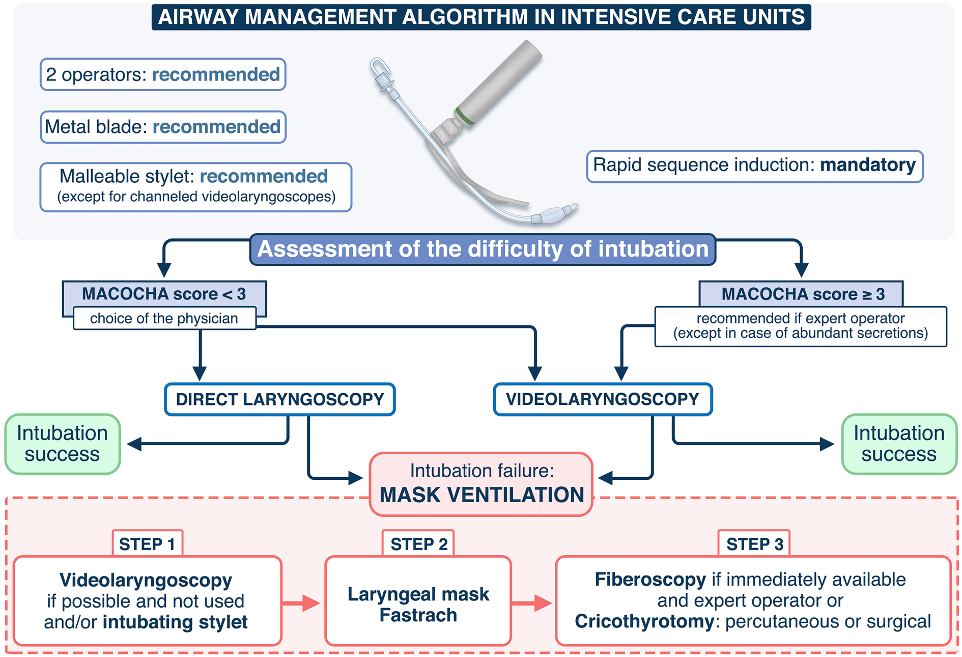
Fig. 1 Airway management algorithm. The availability of equipment for management of a difficult airway is checked. During the procedure, the patient should be ventilated in case of desaturation < 90%. All the intubation procedures performed in ICU are complicated. To improve first-attempt success, two operators, the use of a metal blade and the use of a malleable stylet (except for channeled videolaryngoscopes) are recommended. A rapid sequence induction is mandatory. In case of predicted difficult intubation (Mallampati score III or IV, OSAS, reduced mobility of cervical spine, limited mouth opening, coma, severe hypoxia, non-anesthesiologist (MACOCHA) score ≥ 3), the use of a videolaryngoscope is recommended if the operator is expert in using it (at least 15 intubations performed using the device), excepted in case of abundant secretions. If the MACOCHA score < 3, the choice of the device is left at the operator discretion (direct laryngoscope or videolaryngoscope). In case of intubation failure, a videolaryngoscope will be used if not used first, and/or an intubating stylet (malleable stylet or long flexible angulated stylet), followed successively using Laryngeal Mask Airway or fastrach, the use of fiberscopy in expert hands and finally the use of rescue percutaneal or surgical airway
The use of dedicated tracheal intubation algorithms for critically ill patients [38] in case of predicted or unpre-dicted difficult intubation may also beneficial. We pro-pose an updated airway management algorithm based on recently published trials [54, 55] in Fig. 1. Studies are needed to assess if applying this algorithm in ICU allows reduction of difficult intubation and complications. In each ICU, this airway management algorithm could be adapted according to the needs and situation.
Confirmation of tracheal tube position
Unrecognized esophageal intubation may result in pro-found hypoxemia, brain injury and death [74]. The 4th National Audit Project of the Royal College of Anaesthe-tists and Difficult Airway Society [75] showed that lack of
capnography use contributed to 74% of the airway related deaths in ICU. Hence, clinical examination alone should not be used to exclude esophageal intubation. Following each tracheal intubation, tracheal tube position should be confirmed using continuous sustained waveform capnog-raphy of at least 5–7 breaths [48], even in the case of car-diopulmonary resuscitation. The presence of consistent waveforms reinforces tracheal placement of the endotra-cheal tube, and the effectiveness of cardiopulmonary resuscitation can be monitored by consistently produc-ing EtCO2 values greater than 10–20 mmHg [76]. Inabil-ity to detect sustained exhaled carbon dioxide should prompt immediate laryngoscopic or bronchoscopic con-firmation of tracheal tube position. Tube removal should
1293
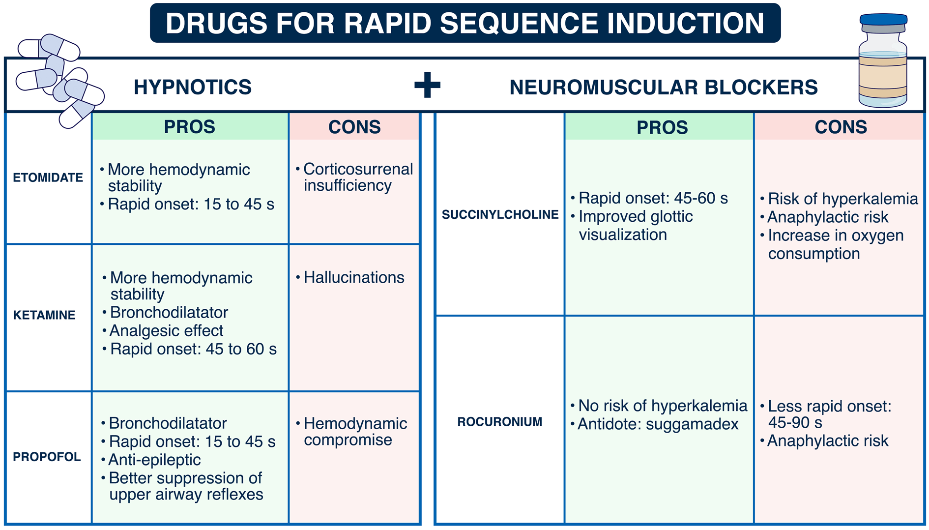
Fig. 2 Drugs used for the intubation procedure: pros and cons
be undertaken with ventilation using a facemask or a supraglottic airway, if esophageal placement cannot be excluded.
Hemodynamic optimization and choice of drugs
Hemodynamic failure is one of the most severe compli-cations associated with endotracheal intubation in the critically ill patients [77]. Peri-intubation cardiovascular collapse is associated with an increased risk of both ICU and 28-day mortality [78]. To prevent severe collapse, fluid loading and early introduction of vasopressors together may decrease the occurrence of hemodynamic intubation-related complications [39, 79]. However, the level of evidence remains low. In a pragmatic, mul-ticenter, unblinded, randomized trial [80], 337 critically ill adults patients undergoing tracheal intubation, were randomly assigned to receive either an intravenous bolus of crystalloid solution only or no fluid bolus. Adminis-tration of an intravenous fluid bolus alone without sys-tematic administration of vasopressors did not decrease the incidence of cardiovascular collapse during tracheal intubation as compared to no fluid bolus. It is worth not-ing that the amount of fluid given was very low, which can partially explain the results, and that it was not com-bined with systematic vasoactive support. Recently, a randomized controlled trial enrolling 1067 critically ill patients undergoing tracheal intubation [81] reported
that administration of an intravenous fluid bolus alone without associated to a systematic administration of nor-epinephrine compared with no fluid bolus did not signifi-cantly decrease the incidence of cardiovascular collapse. The FLUVA trial (NCT05318066) is currently underway to assess the effect of fluid loading and introduction of vasopressors before the tracheal intubation to reduce severe cardiovascular collapse.
The drugs used for intubation [82] are especially important when dealing with hemodynamic complica-tions. After a period of maximal activation of the sympa-thomimetic system all anesthetic drugs will rapidly lead to hemodynamic instability after induction. Vasopressors should be largely used in a preventive way. The respec-tive advantages and benefits of drugs used for intubation are presented in Fig. 2. Russoto et al. [6] recently warned us about the risks of hemodynamic complications using propofol in a post hoc analysis [78] of the INTUBE study. Importantly, these hemodynamic complications were associated with an increased risk of death. Surpris-ingly, rapid sequence induction, combining the use of a neuromuscular blocker and a rapid-onset hypnotic, was used in only 75% of cases [78]. Among patients under-going endotracheal intubation in an out-of-hospital emergency setting, rocuronium, compared with succinyl-choline, failed to demonstrate noninferiority with regard to first-attempt intubation success rate [42]. However, the
1294
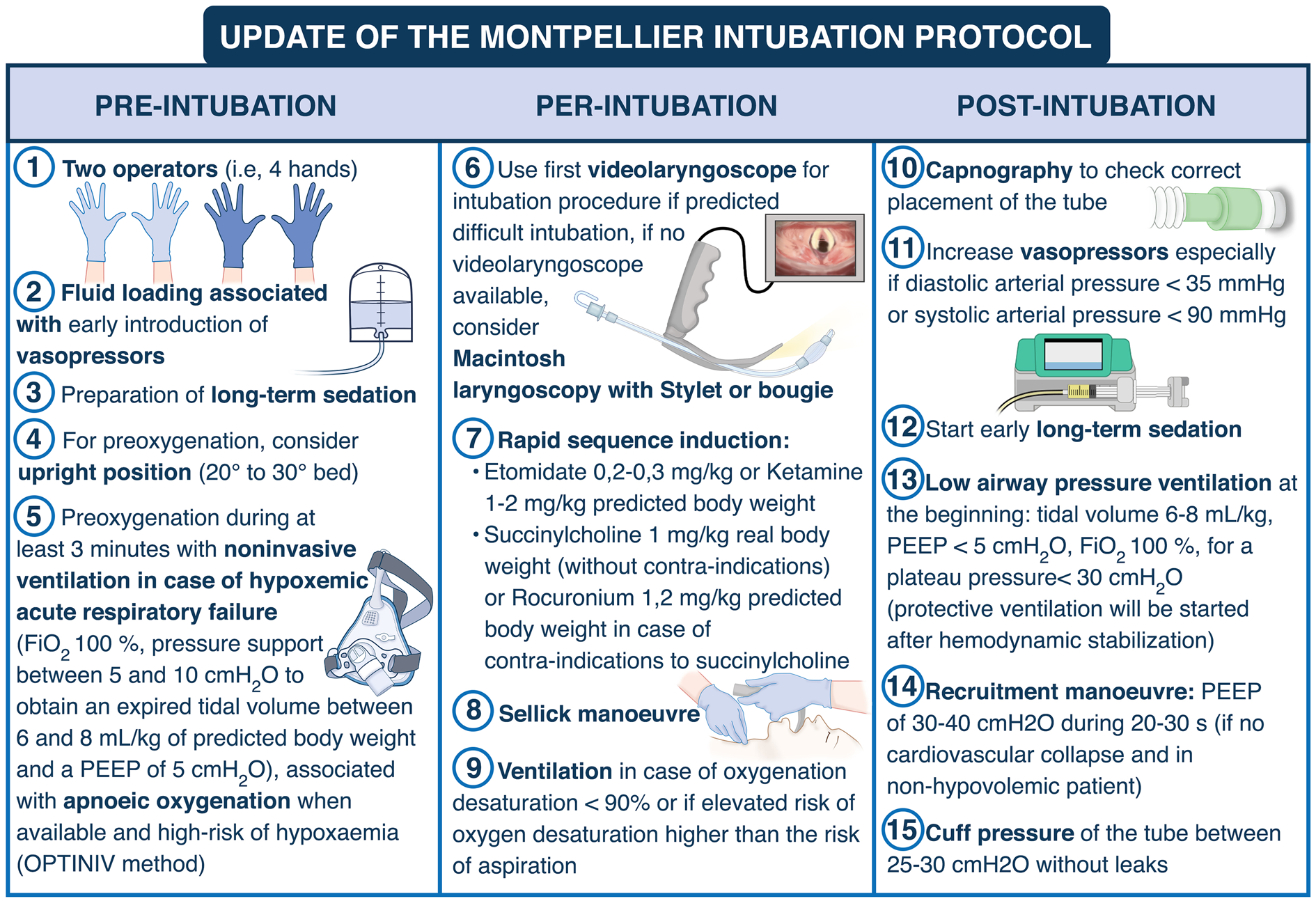
Fig. 3 Update of the Montpellier intubation protocol. Briefly, pre-intubation period interventions consist in fluid loading associated with early introduction of vasopressors, preoxygenation with NIV in the case of acute respiratory failure, preparation of sedation by the nursing team and the presence of two operators. NIV is applied during the 3-min preoxygenation phase with an ICU ventilator and a standard face mask. The PSV level is set between 5 and 10 cmH2O, adjusted to obtain an expired tidal volume of 6 to 8 ml/kg of ideal body weight. The FiO2 is set at 100% and the PEEP level of 5 cmH2O. During the intubation period, recommended induction is rapid sequence induction using short acting, well-tolerated hypnotics (etomidate or ketamine), and a rapid-onset muscle relaxant (succinylcholine or rocuronium), with application of cricoid pressure (Sellick maneu-ver). The Sellick maneuver is performed to prevent gastric contents from leaking into the pharynx, by external obstruction of the esophagus, and associated inhalation of substances into the lungs, as well as vomiting into an unprotected airway. Just after the intubation (post-intubation period), we recommend verification of the tube’s position by capnography (a technique which allows to confirm the endotracheal position of the tube and to verify the absence of esophageal placement), initiation of long-term sedation as soon as possible (to avoid agitation) and use of “protective” mechanical ventilation settings, as defined by the ARDS network
differences between the two drugs were clinically not sig-nificant, suggesting that both drugs can be used safely.
Intubation bundle to limit complications
related to intubation procedure (update of the
Montpellier‑ICU intubation algorithm)
Jaber et al. [39] developed an intubation protocol designed to provide a practical tool that help with plan-ning and optimizing the procedure of intubation. The updated version of the Montpellier intubation proto-col [39], presented in Fig. 3, comprises of a list of items required, things to be done, or points to be consid-ered during each of the phases of tracheal intubation: pre-intubation, per-intubation and post-intubation
[83]. Application of this bundle has demonstrated improved safety during tracheal intubation [39]. In this study, Jaber et al. [39] demonstrated that the use of the Montpellier intubation protocol in the inter-vention phase was associated with significant diminu-tion in life-threatening complications (21% vs. 34%, p= 0.03) and other complications (9 vs. 21%, p= 0.01) compared to the control phase. An external validation of the Montpellier intubation protocol, using a modi-fied version of the protocol, was then performed and published in 2018 by Corl et al. [84]. They found that a modified Montpellier protocol was associated with a significant 16.2% [95% CI 5.1–30.0%] increase in first-attempt intubation success and a 12.6% [95% CI
1295
1.2–23.6%] reduction in all intubation-related compli-cations. Similar to these two studies, in the recent STY-LETO trial performed by Jaber et al. [54], only 25% of patients had a severe complication, which is lower than the rate reported by Russoto et al. in their large obser-vational international study [6].One explanation could be that in the STYLETO study [54], applying the Mont-pellier intubation protocol was highly recommended, which may explain the relatively low rate of complica-tions observed in both groups (endotracheal tube + sty-let and endotracheal tube alone), compared to the rate observed in the INTUBE study [6]. For example, in the INTUBE study [6], capnography was used in only 25% of cases.
The combination of a limited physiologic reserve in the critically ill patients and the potential for difficult mask ventilation and intubation [45] mandates care-ful planning and justifies the use of an algorithmic approach to tracheal intubation, though the benefits of implementation need to be further evaluated.
A randomized controlled trial failed to show superi-ority of a verbal checklist prior to intubation in reduc-ing lower arterial pressure or saturation during the intubation procedure [85]. It is worth noting that this checklist lacked interventions aimed at improving physiological parameters, such as adequate preoxy-genation, fluid load, and vasopressors. Moreover, with the prior significant expertise in airway management in the participating centers and the use of checklists for other ICU procedures, a high penetrance of checklist items may have been present in the control group. Nev-ertheless, a pre-intubation checklist may be effective with less experienced teams and where the checklist includes interventions to optimize physiology [86].
Author details
1 Intensive Care Unit, Anesthesia and Critical Care, Department (DAR‑B), Saint
Eloi, Saint Eloi Teaching Hospital, University of Montpellier, Research Unit: Phy-
MedExp, INSERM U-1046, CNRS, 1, 80 avenue Augustin Fliche, 34295 Montpel-
lier, Cedex 5, France. 2 Department of Anaesthesiology, Critical Care and Pain,
Tata Memorial Hospital, Homi Bhabha National Institute, Mumbai, Maharash-
tra, India. 3 Servei de Medicina Intensiva, Parc TAulí Hospital Universitari, Parc
del Taulí 1, 08028 Sabadell, Spain. 4 Centro de Investigación Biomédica en Red
de Enfermedades Respiratorias (CibeRes), Instituto de Salud Carlos III, Madrid,
Spain.
Publisher’s Note
Springer Nature remains neutral with regard to jurisdictional claims in pub-lished maps and institutional affiliations.
Springer Nature or its licensor holds exclusive rights to this article under a publishing agreement with the author(s) or other rightsholder(s); author self-archiving of the accepted manuscript version of this article is solely governed by the terms of such publishing agreement and applicable law.
Received: 31 May 2022 Accepted: 29 July 2022 Published: 20 August 2022
References
Mercat A, Bernardin G, Combes A, Chiche JD, Diehl JL, du Cheyron D, L’Her E, Perrotin D, Schneider F, Thuong M, Wolff M, Zeni F, Dreyfuss D, Ricard JD (2014) Acquiring procedural skills in ICUs: a prospective multi- center study*. Crit Care Med 42:886–895
5.
De Jong A, Molinari N, Terzi N, Mongardon N, Arnal JM, Guitton C, Allaouchiche B, Paugam-Burtz C, Constantin JM, Lefrant JY, Leone M, Papazian L, Asehnoune K, Maziers N, Azoulay E, Pradel G, Jung B, Jaber S (2013) Early identification of patients at risk for difficult intubation in the intensive care unit: development and validation of the MACOCHA score in a multicenter cohort study. Am J Respir Crit Care Med 187:832–839 Russotto V, Myatra SN, Laffey JG, Tassistro E, Antolini L, Bauer P, Lascarrou 6.
JB, Szuldrzynski K, Camporota L, Pelosi P, Sorbello M, Higgs A, Greif R, Putensen C, Agvald-Öhman C, Chalkias A, Bokums K, Brewster D, Rossi E, Fumagalli R, Pesenti A, Foti G, Bellani G (2021) Intubation practices and adverse peri-intubation events in critically ill patients from 29 countries.
JAMA 325:1164–1172
tory complications of anaesthesia: an analysis of claims against the NHS in England 1995–2007. Anaesthesia 65:556–563
Declarations 11. Grant MJ, Booth A (2009) A typology of reviews: an analysis of 14 review
types and associated methodologies. Health Info Libr J 26:91–108
Conflicts of interest
SJ Jaber reports receiving consulting fees from Drager, Medtronic, Mindray, Fresenius, Baxter, and Fisher & Paykel. ADJ reports receiving remuneration for presentations from Medtronic, Drager and Fisher & Paykel. OR reports receiv-ing research grant from Hamilton Medical AG, speaker fees from Hamilton Medical AG, Fisher & Paykel, Aerogen Ltd and Ambu, and non-financial research support from Timpel. No potential conflict of interest relevant to this article was reported for the other authors.
1296
improves preoxygenation before intubation of hypoxic patients. Am J respiratory support strategy in adults: a clinical practice guideline. Inten-
Respir Crit Care Med 174:171–177 sive Care Med 46:2226–2237
tion and care of respiratory failure in obese patients. Lancet Respir Med 33. Frat JP, Ricard JD, Quenot JP, Pichon N, Demoule A, Forel JM, Mira JP,
4:407–418 Coudroy R, Berquier G, Voisin B, Colin G, Pons B, Danin PE, Devaquet J,
Prat G, Clere-Jehl R, Petitpas F, Vivier E, Razazi K, Nay MA, Souday V, Del-lamonica J, Argaud L, Ehrmann S, Gibelin A, Girault C, Andreu P, Vignon P, Dangers L, Ragot S, Thille AW (2019) Non-invasive ventilation versus high-flow nasal cannula oxygen therapy with apnoeic oxygenation for preoxygenation before intubation of patients with acute hypoxaemic respiratory failure: a randomised, multicentre, open-label trial. Lancet
tive pressure ventilation to enhance preoxygenation in morbidly obese Respir Med 7:303–312
patients: a randomized controlled study. Anesth Analg 107:1707–1713 19. Futier E, Constantin JM, Pelosi P, Chanques G, Massone A, Petit A, Kwiat- kowski F, Bazin JE, Jaber S (2011) Noninvasive ventilation and alveolar recruitment maneuver improve respiratory function during and after intubation of morbidly obese patients: a randomized controlled study.
Anesthesiology 114:1354–1363
Intensive Care Med 42:1336–1349
applications of high-flow nasal cannula in the operating RoomHigh flow nasal cannula. Springer, Cham, pp 101–108
Anesthesiology 20:789–798
improve our practice. Crit Care 18:209
193:273–280 with difficult airways undergoing emergency intubation: a randomized
clinical trial. JAMA 319:2179–2189
45:532–534 sequence intubation: a randomized clinical trial. JAMA 322:2303–2312
endotracheal intubation in the critically ill patient? Pro. Intensive Care success is associated with fewer complications related to intubation in
Med 45:529–531 the intensive care unit. Intensive Care Med 46:1278–1280
for endotracheal intubation in the critically ill patient? Con. Intensive Care Cook TM (2018) Guidelines for the management of tracheal intubation in
Med 45:526–528 critically ill adults. Br J Anaesth 120:323–352
1297
E, Mekontso-Dessap A, Michel F, Nolent P, Perbet S, Prat G, Roquilly A, Tazarourte K, Terzi N, Thille AW, Alves M, Gayat E, Donetti L (2017) Intuba-tion and extubation of the ICU patient. Anaesthesia Crit Care Pain Med
36:327–341 63. De Jong A, Sfara T, Pouzeratte Y, Pensier J, Rolle A, Chanques G, Jaber S
(2022) A videolaryngoscope as a first-intention device for tracheal intuba- tion in unselected patients in the operating room. Br J Anaesth.
21:146–153 versus endotracheal tube alone on successful first-attempt tracheal intu-
bation among critically ill patients: the multicentre randomised STYLETO study protocol. BMJ Open 10:e036718
130–139 (2015) Defining and developing expertise in tracheal intubation using
a GlideScope((R)) for anaesthetists with expertise in Macintosh direct laryngoscopy: an in-vivo longitudinal study. Anaesthesia 70:290–295 68. Amalric M, Larcher R, Brunot V, Garnier F, De Jong A, Moulaire Rigollet V, Corne P, Klouche K, Jung B (2020) Impact of videolaryngoscopy expertise on first-attempt intubation success in critically ill patients. Crit Care Med
Mendez-Tellez PA, Hillel AT, Best SR, Levy MJ (2020) Laryngeal injury and 48:e889–e896
upper airway symptoms after endotracheal intubation during surgery: a systematic review and meta-analysis. Anesth Analg 134:1023–1032 54. Jaber S, Rollé A, Godet T, Terzi N, Riu B, Asfar P, Bourenne J, Ramin S, Lemi- ale V, Quenot JP, Guitton C, Prudhomme E, Quemeneur C, Blondonnet R, Biais M, Muller L, Ouattara A, Ferrandiere M, Saint-Léger P, Rimmelé T, Pottecher J, Chanques G, Belafia F, Chauveton C, Huguet H, Asehnoune K, Futier E, Azoulay E, Molinari N, De Jong A (2021) Effect of the use of an endotracheal tube and stylet versus an endotracheal tube alone on first- attempt intubation success: a multicentre, randomised clinical trial in 999 patients. Intensive Care Med 47:653–664
direct laryngoscopy for adult surgical and intensive care unit patients requiring tracheal intubation: a systematic review and meta-analysis of randomized controlled trials. Eur Rev Med Pharmacol Sci 25:7734–7749 70. Cook TM, El-Boghdadly K, McGuire B, McNarry AF, Patel A, Higgs A
(2020) Consensus guidelines for managing the airway in patients with COVID-19: Guidelines from the Difficult Airway Society, the Association of Anaesthetists the Intensive Care Society, the Faculty of Intensive Care Medicine and the Royal College of Anaesthetists. Anaesthesia 75:785–799 71. Patwa A, Shah A, Garg R, Divatia JV, Kundra P, Doctor JR, Shetty SR, Ahmed SM, Das S, Myatra SN (2020) All India difficult airway association (AIDAA) consensus guidelines for airway management in the operating room during the COVID-19 pandemic. Indian J Anaesth 64:S107-s115
326:2488–2497 Dahyot-Fizelier C, Dahmani S, de Queiroz M, Di Maria S, Ecoffey C, Futier E,
Geeraerts T, Jaber H, Heyer L, Hoteit R, Joannes-Boyau O, Kern D, Lange- ron O, Lasocki S, Launey Y, le Saché F, Lukaszewicz AC, Maurice-Szambur- ski A, Mayeur N, Michel F, Minville V, Mirek S, Montravers P, Morau E, Muller L, Muret J, Nouette-Gaulain K, Orban JC, Orliaguet G, Perrigault PF, Plantet F, Pottecher J, Quesnel C, Reubrecht V, Rozec B, Tavernier B, Veber B, Veyckmans F, Charbonneau H, Constant I, Frasca D, Fischer MO, Huraux C, Blet A, Garnier M (2020) Guidelines: Anaesthesia in the context of COVID- 19 pandemic. Anaesthesia, critical care & pain medicine 39:395–415 74. Holland R, Webb RK, Runciman WB (1993) The Australian Incident
Monitoring Study. Oesophageal intubation: an analysis of 2000 incident reports. Anaesth Intensive Care 21:608–610
a meta-analysis of randomised trials. Intensive Care Med 43:947–948 7:332–340
1298
A, Greif R, Pesenti A, Valsecchi MG, Fumagalli R, Foti G, Bellani G, Laffey JG (2022) Peri-intubation cardiovascular collapse in critically ill patients: insights from the INTUBE study. Am J Respir Crit Care Med.
(2015) Incidence of and risk factors for severe cardiovascular collapse after endotracheal intubation in the ICU: a multicenter observational study. Crit Care 19:257
AN, Gulati S, Stigler WS, Bentov I, Joffe AM, Rice TW (2019) Effect of a fluid
bolus on cardiovascular collapse among critically ill adults undergoing 84. Corl KA, Dado C, Agarwal A, Azab N, Amass T, Marks SJ, Levy MM, Mer-
tracheal intubation (PrePARE): a randomised controlled trial. Lancet Respir chant RC, Aliotta J (2018) A modified Montpellier protocol for intubating
Med 7:1039–1047 intensive care unit patients is associated with an increase in first-pass
intubation success and fewer complications. J Crit Care 44:191–195 85. Janz DR, Semler MW, Joffe AM, Casey JD, Lentz RJ, de Boisblanc BP, Khan YA, Santanilla JI, Bentov I, Rice TW (2017) A multicenter, randomized trial of a checklist for endotracheal intubation of critically ill adults. CHEST J 153(4):816–824.
Referências
Conteúdo migrado do Central de Estudos (Blog Dr. Jackson Fuck, Notion).