Muscle weakness after critical illness: unravelling biologic
Visão geral
https://ccforum.biomedcentral.com/articles/10.1186/s13054-025-05462-z
📋 RESUMO DETALHADO E DIDÁTICO
🎯 CONTEXTO E PROBLEMA PRINCIPAL
O número de sobreviventes de UTI está aumentando devido aos avanços no cuidado hospitalar, chegando a aproximadamente 16 milhões de pessoas por ano no mundo. No entanto, esses sobreviventes enfrentam consequências graves e duradouras conhecidas como Síndrome Pós-Cuidados Intensivos (PICS).
A principal característica dessa síndrome é a fraqueza muscular persistente, que pode durar anos após a alta da UTI e resulta em:
- Redução da capacidade de exercício
- Incapacidade física prolongada
- Maior risco de mortalidade (até 5 anos após alta)
- Custos elevados com saúde e reabilitação
🔬 LACUNA NO CONHECIMENTO
Apesar de décadas de pesquisa sobre a fraqueza muscular adquirida na UTI, os mecanismos subjacentes da disfunção muscular pós-UTI ainda são mal compreendidos. Não existe tratamento baseado em evidências para melhorar os resultados físicos a longo prazo.
Hipótese central: A fisiopatologia é dinâmica ao longo do tempo, sendo diferente entre o período agudo na UTI e a fase de recuperação após a alta.
📊 CONSEQUÊNCIAS FÍSICAS A LONGO PRAZO
Durante a UTI:
A fraqueza adquirida na UTI (ICUAW) é definida clinicamente por uma pontuação MRC (Medical Research Council) < 48, sem causa aparente além da doença crítica.
Após a alta da UTI:
- Primeiros 3-6 meses: Melhora inicial nas capacidades físicas
- Após 6 meses: A função muscular atinge um platô que persiste por anos
- Prevalência: Mais de 60% dos sobreviventes de sepse apresentam fraqueza persistente; mais de 90% têm comprometimentos físicos prolongados
- Primeiro ano: Quedas significativas na força muscular máxima e de resistência
- Até 5 anos: Limitações na função física persistem, com redução na capacidade aeróbica (24% de redução no VO2 pico comparado a controles saudáveis)
- Autonomia: Quase metade dos pacientes que viviam independentemente antes da hospitalização perdem essa capacidade 6 meses após a UTI
⚕️ FATORES CLÍNICOS QUE INFLUENCIAM A RECUPERAÇÃO
🔹 Status de Saúde Pré-UTI:
Idade e Sexo
Idade: Indivíduos mais velhos têm piores resultados a longo prazo
Sexo: Mulheres são mais propensas à ICUAW e apresentam maior comprometimento da função física comparadas aos homens
Causas possíveis em mulheres: Redução de miofibras tipo IIa, menor sensibilidade à insulina, deficiência de estrogênio
Obesidade Pré-mórbida
Obesidade Pré-mórbida
Efeito protetor: A obesidade atenua a taxa de mortalidade e fraqueza muscular
Mecanismo: Mobilização mais eficiente de ácidos graxos endógenos e maior disponibilidade de corpos cetônicos, prevenindo o desgaste do tecido magro
- Efeito independente do estado nutricional ou atividade da leptina
Sarcopenia Pré-existente
Sarcopenia Pré-existente
Definição: Perda de massa, força e desempenho muscular devido ao envelhecimento (primária) ou doenças subjacentes como câncer (secundária)
Impacto: Fator agravante para fraqueza muscular pós-UTI, mesmo após ajuste por idade e comorbidades
- Explica melhor o status funcional de longo prazo do que a persistência da perda de massa muscular após 3 meses
🔹 Durante a Permanência na UTI:
Gravidade da Doença
Gravidade da Doença
Principais causas: Síndrome do Desconforto Respiratório Agudo (SDRA) e choque séptico
Duração do suporte orgânico artificial: Especialmente ventilação mecânica
Tempo de permanência na UTI: Associado à má recuperação física
Doença Crítica Crônica (DCC): Disfunção orgânica prolongada (>10 dias) com inflamação, imunossupressão e síndrome de catabolismo persistentes
Hiperglicemia e Nutrição
Hiperglicemia e Nutrição
Hiperglicemia: Fator de risco conhecido para ICUAW; pode afetar função mitocondrial e autofagia
Déficit calórico: Relatado como fator de risco para desgaste muscular, mas estudos mostram resultados contraditórios
Alimentação precoce completa: Não melhora resultados funcionais de longo prazo; pode piorar hiperglicemia e disfunção de autofagia
Estratégia sugerida: Restrição calórica e proteica na fase aguda (primeiros 7 dias) leva a recuperação mais rápida
Imobilidade e Medicamentos
Imobilidade e Medicamentos
Imobilidade: Repouso prolongado no leito e inatividade levam à atrofia muscular rápida
Corticosteroides e bloqueadores neuromusculares (BNM): Impacto controverso; pode depender da duração, dose e indicação
Recomendação: Uso adequado de medicamentos, minimizando dose de corticosteroides
🔹 Status de Saúde Pós-UTI:
Fraqueza muscular na alta da UTI: Forte preditor de má recuperação física
Importância: Avaliar a pontuação MRC antes da alta hospitalar para identificar pacientes de alto risco precocemente
🧬 MECANISMOS BIOLÓGICOS (Visão Geral)
A revisão aborda mecanismos complexos e multifacetados que impulsionam a disfunção muscular pós-UTI:
🎯 OBJETIVOS DA REVISÃO
Esta revisão narrativa visa abordar os determinantes clínicos, fisiológicos e biológicos da disfunção muscular persistente em sobreviventes de UTI, com atenção especial aos mecanismos moleculares, celulares e sistêmicos envolvidos.
A revisão também:
💡 MENSAGEM-CHAVE
Abstract
Survivors of intensive care unit (ICU) are increasingly numerous because of better hospital care. However, several consequences of an ICU stay, known as post-intensive care syndrome, worsen long-term prognoses. A predominant feature in survivors is reduced muscle strength, mass, and physical function. This leads to lower exercise capacity, long-lasting physical disability, higher mortality risk, and subsequent health costs. While ICU-acquired muscle weakness has been extensively studied these past decades, underlying mechanisms of post-ICU muscle weakness remain poorly understood, and there is still no evidence-based treatment for improving long-term physical outcomes. One hypothesis, among others, could be that the pathophysiology is dynamic over time, differing between the acute ICU and post-ICU recovery periods. This narrative review aims to address the clinical, physiological and biological determinants of persistent muscle dysfunction in ICU survivors, with particular attention to the molecular, cellular and systemic mechanisms involved. Specifically, pre-ICU health factors such as obesity and sarcopenia, ICU-related complications and treatments, and post-ICU management all influence recovery. Dysfunctions in the neuroendocrine, vascular, neurological, and muscle systems contribute as physiological determinants of the muscle weakness. Complex and multifaceted biological mechanisms drive the post-ICU muscle dysfunction with mitochondrial and autophagy dysfunction, epigenetic modifications, cellular senescence, muscle inflammation with altered cell–cell communication, including dysfunction of immune cells, stem cell exhaustion and extracellular matrix remodelling. The review also sheds light on new and innovative therapeutic approaches and discusses future research directions. Emphasis is placed on the potential for multi-approach treatments that integrate nutritional, physical, and biological interventions. Addressing these aspects in a holistic and dynamic manner, from ICU to post-ICU phases, may provide avenues for mitigating the long-term burden of muscle weakness and physical disability in ICU survivors.
Introduction
Despite the increasing age and severity of illness among Intensive Care Unit (ICU) patients, the number of survivors is substantial and rising, reaching approximately 16 million worldwide each year because of better hospital care [1]. However, several long-lasting consequences after ICU stay, known as post-intensive care syndrome (PICS), impair long-term prognoses. A predominant feature in survivors is the reduction of muscle strength during their hospital stay that persists years after ICU discharge and results in reduced exercise capacity, long-lasting physical disability, distress, and higher subsequent mortality risk up to 5 years after discharge [2,3,4]. It also results in further health costs, as many survivors require rehabilitation and long-term facilities, meaning that PICS is becoming a significant public health challenge [5].
Despite numerous studies describing the long-term ICU consequences on skeletal muscle and physical function, research exploring the physiological and biological mechanisms remains scarce and there is still no evidence-based treatment for improving long-term outcomes. One hypothesis, among others, could be that the pathophysiology is dynamic over time, differing between the acute ICU period and the recovery phase after ICU discharge. In this field, the most significant knowledge is based on experimental models and ICU-surviving patients who experienced sepsis, acute lung injury and mechanical ventilation.
This review provides an integrative and updated perspective on post-ICU muscle dysfunction, going beyond prior work by bridging clinical, physiological, and molecular insights across the ICU and post-ICU continuum. By framing muscle weakness as a dynamic and multifactorial process, the review offers a comprehensive foundation for future therapeutic strategies. The detailed methodology used to conduct this review is described in the supplementary materials (see Additional file 1).
Long-term muscle and physical consequences
While ICU-acquired weakness (ICUAW), clinically defined by a Medical Research Council (MRC) sum score < 48 in patients with no plausible cause other than critical illness, worsens the short-term consequences (e.g., increased mechanical ventilation duration, length of stay, and in-hospital mortality), it also impacts long-term outcomes [1, 6]. It is well known that after an initial improvement in physical capacities during the first three to six months following ICU discharge, muscle function in survivors reaches a plateau that persists for years thereafter [7]. Long-term studies on ICU survivors indicate that these individuals often suffer from persistent weakness and prolonged physical impairments, with reported prevalence exceeding 60% and 90%, respectively, among sepsis survivors [8]. Within the first year after ICU discharge, survivors exhibit significant declines in muscle maximal and endurance forces [2]. Muscle weakness directly and negatively impacts physical performance and health-related quality of life (QOL) [9]. Physical functioning limitations persist for up to five years, as evidenced by reduced distance in the 6-min walk test and aerobic capacity [4, 10]. Indeed, during the 5-year follow-up of the EPaNIC study, 361 patients underwent cardiopulmonary exercise testing, which revealed a 24% reduction in peak VO2 compared with healthy controls. This deterioration in exercise capacity involved muscle limitations in more than 60% of cases [4]. The decline in physical ability can severely impact patient autonomy, with nearly half of those living independently before hospitalization losing this ability six months post-ICU [11]. However, recovery trajectories vary among ICU survivors [12], and while some ICU survivors may experience improvements in functional status, others may never achieve full recovery. Indeed, two-thirds of septic shock survivors had not regained their pre-ICU physical status one year after discharge [10]. The physical recovery is influenced by clinical, physiological, and biological determinants, with long-lasting molecular, cellular, and systemic abnormalities.
Clinical factors
Pre-ICU health status
Similar to its impact on short-term ICU outcomes, age also affects long-term physical recovery, with older individuals experiencing poorer outcomes [13]. While females are more prone to ICUAW, they exhibit greater impairment of physical function in the long term compared with males [14, 15]. Reduced type IIa myofibers, decreased insulin sensitivity and oestrogen deficiency in critically ill females have been suggested as contributing factors to the observed gender disparities [16,17,18]. Premorbid obesity is a well-documented factor that attenuates the mortality rate and muscle weakness in both humans and animals [19]. Its effects are independent of the nutritional status (fasting or parenteral feeding) or the activity of the leptin adipokine [20, 21]. It appears to be related to a more efficient mobilization of endogenous fatty acids and an increased availability of ketone bodies, preventing lean tissue wasting [20, 22]. Conversely, pre-existing sarcopenia, defined by the loss of muscle mass, strength, and physical performance due to aging (primary sarcopenia) or underlying diseases (secondary sarcopenia, such as cancer), is a worsening factor for post-ICU muscle weakness, even after adjusting for age and comorbidity levels [23, 24]. This condition better explains the poor long-term functional status than the persistence of muscle mass loss at 3 months after sepsis [23]. Unravelling the causal mechanisms of pre-ICU health status may facilitate the development of targeted strategies (Fig. 1).
Clinical factors influencing the post-ICU physical recovery. ARDS: acute respiratory distress syndrome, ICU: Intensive Care Unit, ICUAW: ICU acquired weakness, NMBA: neuromuscular blocking agents, QOF: quality of lifeFull size image
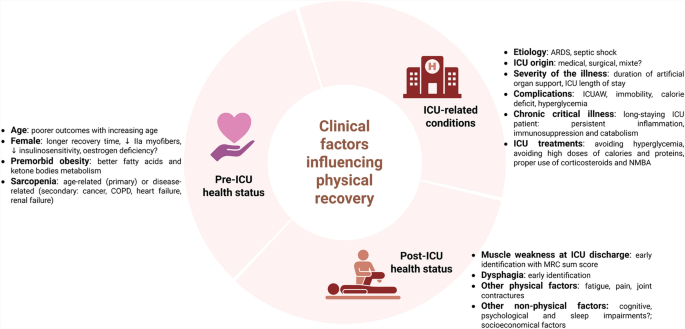
The extent of physical impairment is closely linked to the severity of the illness upon admission and the reason for hospitalization, particularly acute respiratory distress syndrome (ARDS) and septic shock [9, 25, 26] (Fig. 1). The duration of artificial organ support, especially mechanical ventilation, and the length of ICU stay are both associated with poor physical recovery [27, 28]. Importantly, Chronic Critical Illness (CCI), defined by prolonged organ dysfunction lasting more than ten days, has a significant impact on long-term physical function [29]. These long-staying ICU patients frequently suffer from persistent inflammation, immunosuppression, and catabolism syndrome, which further complexifies the pathophysiology [28]. The potential influence of ICU admission origin — surgical vs. medical — on post-ICU muscle dysfunction have not been specifically compared. Most available data come from mixed cohorts, which may introduce heterogeneity and limit the interpretation of findings. Future studies are warranted to determine whether distinct processes contribute to muscle dysfunction in surgical versus medical populations (See Additional file 2).
Hyperglycaemia, a well-known risk factor for ICU-acquired weakness (ICUAW) [6], and its treatment with insulin may disrupt neuronal and muscle cell homeostasis by affecting mitochondrial function and autophagy [30]. However, the role of hyperglycaemia in the ICU as either a pathogenic or adaptive response remains debated. Calorie deficit – constituted in the acute phase – is reported to be a risk factor for muscle wasting in ICU survivors, independent of the severity of illness [31]. Nevertheless, early instauration of full feeding does not improve long-term functional outcomes and these patients experience slower physical recovery [32,33,34]. This may be attributed to anabolism resistance, worsening of hyperglycaemia, insulin needs, and autophagy dysfunction [35, 36]. Conversely, experimental studies demonstrate that the energy deficit does not worsen the muscle phenotype in ICU animals [35, 37,38,39]. In line with these results, restricting calorie and protein intake (6 kcal/kg/d and 0.2–0.4 g/kg/d versus 25 kcal/kg/d and 1.0–1.3 g/kg/d) in the acute phase (i.e. first 7 days) leads to a faster recovery without harmful effects in critically ill patients [40]. Immobility due to prolonged bed rest and inactivity leads to rapid muscle atrophy and exacerbates neuromuscular dysfunction [6]. Studies of the impact of corticosteroids and neuromuscular blocking agents (NMBA) on physical recovery are controversial and may depend on the administration duration, dose, and indication [9, 27, 41, 42]. These data support the proper use of medicines, including minimizing the dose of corticosteroids (Fig. 1).
Post-ICU health status
Muscle weakness at ICU discharge is a strong predictor of poor physical recovery, underscoring the need to identify high-risk patients early. Assessing the MRC sum score remains crucial before hospital discharge. Survivors with a low MRC score at ICU discharge, particularly those scoring below 55, have a higher risk of long-term mortality and poor outcomes [1, 3, 6]. Importantly, MRC scores at ICU discharge correlate with long-term measures of muscle strength, physical function, and quality of life, even five years after ICU discharge [3]. In addition to peripheral muscle weakness, ICU-acquired dysphagia, negatively influences physical health [43]. Approximately 30% of mechanically ventilated patients develop clinically significant post-extubation dysphagia (PED), which may persist in up to 18% of sepsis survivors three years after ICU discharge [8, 44]. The pathophysiology of PED is multifactorial, encompassing local laryngo-pharyngeal trauma induced by endotracheal intubation, pharyngeal and laryngeal muscles dysfunction, and impaired central regulation of swallowing [45]. PED is associated with delayed resumption of oral feeding, increased risk of malnutrition, delayed physical recovery, prolonged hospital stay, and higher mortality [46, 47]. Expert recommendations now advocate for systematic screening and early management of swallowing disorders in critically ill patients, as part of a comprehensive post-ICU rehabilitation strategy aimed at optimizing nutritional status and functional outcomes [45] (Fig. 1).
Putative and established post-ICU clinical factors influencing physical recovery. Factors related to physical health are shown in red (left), mental health in blue (upper right), and other factors in yellow (lower right). PICS: post-intensive care syndromeFull size image
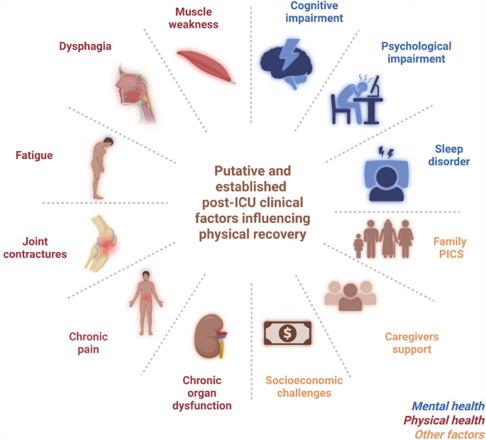
Beyond muscle pathology, survivors of critical illness face numerous barriers that hinder physical recovery. Fatigue, highly prevalent in ICU survivors may impact physical recovery and is addressed in the “Neurological dysfunction” section. Persistent pain is frequently reported in this population, with up to 66% of ICU survivors develop new chronic pain in the months following discharge [8, 48]. Joint contractures affect over one-third of ICU survivors at hospital discharge and further restrict mobility and complicate rehabilitation [49, 50]. Other new chronic organ dysfunction, such as post-ICU chronic kidney failure, negatively modifies long-term functional outcomes [51, 52]. In addition, poor mental health may adversely influence physical recovery. More than 60% of survivors experience sleep disorders, often persisting for months after discharge [53, 54]. Psychological impairments, including depression, anxiety, and post-traumatic stress disorder, and cognitive impairments, including deficits in attention, memory, and executive function, are observed in more than one-third of ICU survivors at one year [55,56,57,58]., Furthermore, ICU survivors face significant socioeconomic challenges, as functional impairments often limit their ability to return to work, leading to substantial lost earnings [59, 60]. Finally, the burden of critical illness extends beyond the patient. Family involvement may be a critical determinant of functional recovery, yet caregivers frequently report limited resources, and a lack of support [61, 62]. Overall, addressing pain control, prevention of contractures, neuropsychological support, family-centered care, and socioeconomic reintegration may improve the long-term physical outcomes in addition to muscle-specific therapies (Fig. 2).
Physiological determinants
Neuroendocrine abnormalities
In the early phase (e.g. first days), impairment in the peripheral hormone metabolism leads to active secretion from the anterior pituitary, reduced availability of anabolic hormones, and increased availability of catabolic hormones [63]. In the late phase (e.g. after a few days), neurohormonal secretions are repressed, primarily due to hypothalamic deficit, with insufficient levels of peripheral hormones such as cortisol [63, 64]. After critical illness, knowledge is scarce. Persistent hypopituitarism lasts for years in patients after traumatic brain injury and is associated with poor physical outcomes and quality of life [65]. In non-brain injured patients, Vanhorebeek et al. explored the somatotropic, thyroid, and adrenal axis 5 years after ICU discharge in the EPaNIC follow-up study [66]. While most acute changes resolve, inactive reverse triiodothyronine (rT3) levels and the T3/rT3 ratio remain low, and are correlated with reduced hand grip strength [66]. Whether targeting the long-term thyroid axis dysfunction may improve physical recovery remains to be demonstrated in translational studies (Fig. 3 and Table 1).
Physiological determinants of the post-ICU muscle weakness at the system level. Further research directions are highlighted in italics. T3: triiodothyronine, rT3: inactive reverse T3, CNS: Central nervous system, PNS: peripheral nervous system, NMJ: neuromuscular junction, RCT: randomized controlled trialFull size image
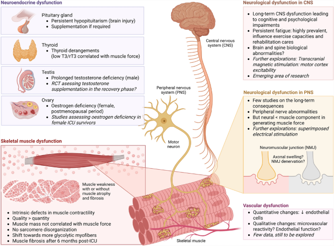
Table 1 Summary of the dynamic pathophysiology changes from the ICU period to the post-ICU convalescence and further research perspectives
Critically ill male patients often experience significant and prolonged testosterone deficiency, which occurs rapidly upon ICU admission and lasts after ICU discharge [67]. Hypogonadism has been associated with low muscle mass and function in ICU patients [17]. Testosterone or analog treatments alleviate muscle catabolism in severely burned patients [68] and improve muscle phenotype in septic animals [69]. Whether testosterone may overcome the anabolic resistance remains to be demonstrated in non-burn ICU survivors. As testosterone or analog treatment appears to be safe [70], RCTs assessing its effect in the recovery phase are warranted.
Female sex is a well-established risk factor for ICUAW and poor physical recovery [14, 15], potentially linked to oestrogen deficiency. Postmenopausal women experience a chronic decline in oestrogen levels, which is associated with accelerated muscle mass and strength loss compared to men [71]. Hormone replacement therapy has been shown to preserve muscle function in this population [18]. Oestrogen deficiency impairs satellite cell activation, promotes muscle fibre atrophy, and delays regeneration after injury [72]. While younger women may benefit from endogenous oestrogen, critical illness itself may transiently suppress gonadal function [17], potentially reducing this protection. Thus, oestrogen deficiency — especially in older female ICU survivors — may represent a relevant but underrecognized factor contributing to long-term muscle weakness [73]. Further investigations are warranted to elucidate the role of oestrogen in post-critical illness physical recovery (Fig. 3 and Table 1).
Vascular dysfunction
Endothelial dysfunction plays a role in the pathogenesis of ICUAW. However, studies have focused on the first hours of critical illness, and little is known about the convalescent phase [74]. Although microvascular dysfunction improved throughout ICU stay [75], microvascular reactivity defects remain at ICU discharge [76]. In sepsis-surviving mice, quantitative changes are observed with a reduced number of endothelial cells [77]. Given the existing endothelial-myocyte crosstalk, further research is required to explore the role of the vascular system in long-term physical recovery (Fig. 3).
Neurological dysfunction
Peripheral nerve injuries may play a role in the pathophysiology of long-lasting muscle weakness, as neuromuscular junction and axonal abnormalities are observed in animal models of prolonged ICU stay [78]. Nevertheless, in situ contractility experiments with both direct muscle stimulation and indirect nerve stimulation indicate that the neural component plays a minimal role in reduced muscle force. Instead, alterations in the muscle component appear as the primary mechanism [78]. In humans, electromyography (EMG) and nerve conduction studies indicate electrophysiological evidence of myopathy in 79% of ICU survivors, with those affected experiencing more severe weakness [79]. Physiological assessment, combining surface EMG and ergometry, suggests that the reduced contractility in human ICU survivors one year after hospitalization originates in muscle tissue rather than the nervous system [2]. Further physiological assessments of the motor control, such as superimposed electrical stimulation and transcranial stimulation, may help in deciphering other neurological mechanisms underlying the muscle weakness (Fig. 3 and Table 1).
Even if muscles and nerves are functionally and structurally intact, the decreased muscle activation by central nervous system (CNS) can lead to muscle weakness and fatigue. Fatigue is defined as “an overwhelming sense of tiredness, lack of energy and feeling of exhaustion, fatigue relates to a difficulty in performing voluntary tasks” [80]. Fatigue is commonly reported by ICU survivors, in more than 57% up to 5 years after discharge, with women more likely to be affected than men [81,82,83,84]. Fatigue is a determinant of physical performance, limits exercise capacity and influences rehabilitation care [85]. While fatigue is prevalent after critical illness, the underlying mechanisms have not been studied. Post-infectious myalgic encephalomyelitis/chronic fatigue syndrome (ME/CFS) is a complex and debilitating disorder following an infection, characterized by fatigue that cannot be explained by any underlying medical condition. Wallit et al. recently demonstrated that patients with ME/CFS exhibited dysfunction in integrative brain regions, decreased parasympathetic activity, and dysregulation of catecholamine pathways, which correlate with reduced muscle strength [86]. While these findings in ME/CFS patients cannot be directly applied to ICU survivors, these data suggest that CNS determinants may play a role in post-sepsis fatigue. In conclusion, much research remains to be done in this field to address significant knowledge gaps that require further investigation [87] (Fig. 3 and Table 1).
Muscle dysfunction
The muscle component of ICUAW is characterized by two key features: (1) altered myofiber contractility, and; (2) myofiber atrophy (reduced myofiber size and number), with the following hallmarks: altered muscle cell excitability, sarcomere disorganization, preferential loss of myosin over actin, and greater loss of MyHc II (fast-twitch fibers) than Myhc I (slow-twitch fibers) – features not observed in simple bedrest conditions [39, 88, 89]. After critical illness, myofiber contractility and atrophy can be dissociated. Indeed, both animal and human studies have shown persistent muscle weakness despite muscle mass restoration in ICU survivors [84, 90, 91]. In addition, Dos Santos et al. found that muscle mass did not correlate with muscle force and even the weakest individuals exhibited restored muscle mass [79]. While sarcomere disorganization and myofiber cross-sectional area is restored in ICU survivors, a shift toward more glycolytic myofibers has been observed [84]. Normalizing muscle force to myofiber cross-sectional area did not alter results, indicating intrinsic contractile dysfunction in ICU survivors, independent of muscle mass. Based on these observations, the biomechanical characteristics of muscle fibres may be at least as important as their quantity (Fig. 3). Contributing mechanisms may include abnormalities in calcium handling, sarcolemma excitability, mitochondrial dysfunction, metabolic imbalances, post-translational modifications in contractile proteins, and fibrosis.
Biological mechanisms
Nuclear and organelle dysfunction
Insulin resistance and impaired glucose metabolism
Insulin resistance is a hallmark of critical illness, triggered by systemic inflammation and stress-induced hormonal changes. Hyperinsulinemic-euglycemic clamp studies have shown up to a 70% reduction in insulin sensitivity in ICU patients, closely associated with illness severity [92]. Impaired insulin signalling leads to defective GLUT4 translocation to the sarcolemma, thereby limiting glucose utilization in skeletal muscle [93]. Insulin resistance may not fully resolve after ICU discharge. ICU survivors often exhibit persistent glucose metabolism alterations, including impaired tolerance and new-onset diabetes (up to 17% at five years, with elevated risk up to five fold) [94,95,96]. Several studies have highlighted the association between insulin sensitivity and muscle outcomes in ICU patients. Weber-Carstens et al. observed that in patients with ICUAW, skeletal muscle exhibited resistance to insulin stimulation, which was associated with blunted muscle glucose uptake and more profound muscle fibre atrophy [93]. Insulin sensitivity in ICU patients correlates positively with muscle strength at awakening and at ICU discharge [97], underscoring the link between metabolic and functional recovery. While muscle activation strategies during the ICU stay, such as protocolized physiotherapy and electrical muscle activation, have shown limited effects on insulin sensitivity [97], the post-ICU period may offer a more favourable window for intervention. As anabolic resistance subside, targeted exercise programs may enhance insulin sensitivity and support functional recovery. Similarly, initiating metformin during early convalescence could help reduce post-ICU insulin resistance and contribute to mitigating persistent metabolic disturbances in the skeletal muscle of ICU survivors [98]. Further studies are needed to explore the metabolic benefits of rehabilitation strategies during the post-critical illness period (Table 1).
Mitochondrial dysfunction
Firstly described in human skeletal muscle during septic shock by Gasparetto et al. in 1983 [99], mitochondrial dysfunction has been particularly studied in critically ill patients since the early 2000s [100]. In 2002, Brealey et al. demonstrated that skeletal muscle energy failure was associated with a poor prognosis and that complex I activity was inversely correlated with the severity of septic shock [101]. The mitochondrial population of skeletal muscle is characterized by reduced mitochondrial biomass and impaired mitochondrial quality control pathway in critical illness [102]. In 2016, Dos Santos et al. investigated this hypothesis, among others, and reported no difference in quantitative mitochondrial content, observed by electron microscopy, in ICU survivors 6 months after discharge compared with age- and sex-matched healthy volunteers. However, the authors did not study mitochondrial ultrastructure or morphology [79]. In these same patients, transcriptomic analysis revealed that mitochondrial-related pathways were dysregulated in the post-ICU period [103]. Mitochondrial-related master micro-RNA (miRNA) regulators influence the transcriptomic response and correlate with muscle outcomes [104]. In 2019, using a murine model of cecal slurry injection, Owen et al. described alterations in mitochondrial ultrastructure, respiration and enzymatic activities up to 1 month after sepsis, which paralleled muscle oxidation and nitrosylation [90]. Up to five years after ICU discharge, the aerobic capacity of patients was reduced mainly due to muscle limitation [4]. Mart et al. showed that human ICU survivors exhibited exercise responses similar to those observed in non-critically ill patients with mitochondrial myopathies. Oxygen utilization measurements were strongly correlated with VO2 peak values and the 6-min walk test, suggesting that the exercise capacity may be limited by oxygen utilization [105]. Recently, Mayer et al. demonstrated mitochondrial alterations in eleven humans 9 months after ICU discharge. Survivors exhibited reduced mitochondrial complex II activity that correlated with decreased exercise capacity and increased fatigue [84]. Finally, no published study to date has directly demonstrated alterations to the function and behaviour of mitochondria in human ICU survivors [106, 107]. Characterizing the mitochondrial population and its quality control pathways using muscle biopsy is warranted in ICU survivors to achieve a comprehensive understanding of the cellular and molecular processes involved. This could further facilitate the development of therapeutic approaches to counteract muscle weakness (Fig. 4 and Table 1).
Biological mechanisms underlying post-ICU muscle weakness at the cellular and molecular levels: nuclear and organelle dysfunction. Further research directions are highlighted in italics. ICU: intensive care unit, UPS: ubiquitin proteasome systemFull size image
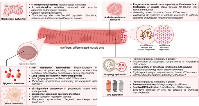
Senescence, is defined as a stable cell-cycle arrest in response to cellular stress, with phenotypic changes such as mitochondrial dysfunction. It can become pathogenic in certain conditions, particularly in survivors of critical care [108, 109]. It leads to the secretion of pro-inflammatory, pro-apoptotic, and pro-fibrotic molecules, known as the senescence-associated secretory phenotype (SASP), perpetuating accelerated aging through low-grade inflammation [110]. While cellular replication has not been studied in the acute phase of critical illness, Chen et al. demonstrated that muscle p53-dependent senescence occurred in sepsis-surviving animals. Importantly, metformin attenuated muscle cellular senescence and loss of muscle strength [107]. Metformin is a well-documented senotherapy acting through mechanisms including AMPK activation, mTOR inhibition, autophagy enhancement, and promotion of mitochondrial biogenesis, resulting in anti-oxidative and anti-inflammatory effects [98]. However, metformin's broad activity in the human body and challenges in its use in critically ill patients suggest targeted therapy may be preferable. In this regard, targeted senolytics — drugs that eliminate senescent cells — were first used in humans in 2019 to improve physical performance [111]. While this research field is rapidly expanding, it holds promise for ICU survivors [108]. Future studies should focus on assessing senescence signalling pathways in the skeletal muscle of ICU survivors to validate the relevance of this hypothesis (Fig. 4 and Table 1).
DNA methylation abnormalities
Epigenetic abnormalities, by disrupting gene expression and cellular phenotype, are linked to human diseases and affect critically ill patients through modifications of the entire epigenetic network [112]. Van Dyck et al. conducted the largest study on the subject and studied the skeletal muscle DNA methylation of 172 critically ill patients in 2022. They identified two hypomethylated regions in the promoters of the HIC1 and NADK2 genes in skeletal muscle, which are crucial for muscle regeneration, postsynaptic acetylcholine receptors, and mitochondrial homeostasis, respectively [113]. While the muscle methylation status was assessed during the ICU stay (day 8 ± 1), no data are available in ICU survivors. Modulation of aberrant DNA methylation and the subsequent transcriptional program may be an innovative approach to alleviate the global, long-term epigenetic issues in ICU survivors (Fig. 4 and Table 1).
Anabolism resistance
Interventions aimed at promoting anabolism to counteract catabolism and restore muscle mass have been examined since 1999. Notably, the first large, double-blind RCT assessing growth hormone in patients with prolonged critical illness reported a two-fold increase in mortality compared with placebo [114], suggesting anabolism resistance. This issue remains relevant two decades later, as Chapple et al. have recently demonstrated the inability of critically ill patients under mechanical ventilation to utilize ingested protein for muscle protein synthesis [115]. Anabolism resistance is now a well-documented phenomenon in critically ill patients, yet it remains unclear whether this condition persists in survivors. In long-staying ICU patients, muscle protein turnover is negative between days 10 and 20 after admission, but the synthesis rate subsequently increases to reach the breakdown rate, achieving an equilibrium in protein balance by days 30 to 40 [116]. Crowell et al. identified the Akt-TSC2-mTORC1 pathway as a key driver of muscle mass recovery in post-septic mice, independently of AMPK activity. Muscle protein synthesis, initially suppressed, doubled during recovery, coinciding with body weight restoration [91, 117]. These data demonstrate that the anabolic resistance progressively fades during the period of recovery. Nevertheless, studies assessing protein turnover in human ICU survivors are currently lacking. Understanding the dynamics of anabolic resistance in humans is essential for clinicians, as the transition from this state toward responsiveness may drive the appropriate refeeding and physical rehabilitation (Fig. 4 and Table 1).
Proteasome dysfunction
The ubiquitin–proteasome system (UPS), intensely activated in critically ill patients, is directly responsible for acute muscle wasting [118]. Experimentally, atrophy-related genes, including E3 ubiquitin ligases, were highly up-regulated in sepsis mice, partly explained by reduced calorie intake [37]. Long-term critical illness led to a necrotizing muscle phenotype linked to proteasomal overload, indicating a build-up of inadequately degraded proteins by the UPS [119]. Additionally, in an experimental extensive-burn model, both early and late pharmacological inhibition of the proteasome with bortezomib reduced the hypermetabolic muscle response but resulted in increased mortality [120]. Although proteolysis seems harmful to muscle mass, its balanced activation remains essential for muscle homeostasis [121]. Finally, while UPS activation and sarcomere destruction were observed in all ICU survivors 7 days post-discharge, these conditions were fully resolved 6 months later [79]. Overall, inhibition of the UPS does not seem to be an effective therapeutic strategy for improving the long-term physical capacity (Fig. 4 and Table 1).
Autophagy dysfunction
Damaged cellular components accumulate during critical illnesses due to inflammation and oxidative stress in the skeletal muscle [1]. Autophagy, a highly evolutionarily conserved process, recycles unnecessary or dysfunctional components through a lysosome-dependent degradation [122]. Nonetheless, the specific role of autophagy in ICU patients remains poorly understood, especially in skeletal muscle [123]. In the acute phase, muscle autophagy is mainly activated through Akt/mTOR inhibition and AMPK activation [123] and may reduce muscle mass. Thus, inhibiting catabolic pathways could prevent muscle wasting in the short term, in preclinical models [124, 125]. However, this does not mean that the overall effect is beneficial on muscle function in the long term. Long-stay ICU patients accumulated up to tenfold more immature or unfused autophagosomes than matched controls, suggesting an insufficient activation of autophagy [126]. Pharmacological or genetic inhibition of autophagy worsened the course of sepsis with increased mortality rate and disease severity [127,128,129]. In the recovery period, the muscle-specific and inducible deletion of Atg7 worsens the muscle outcomes in sepsis-surviving mice [129]. Transcriptomic analyses revealed that it strongly activates other catabolism pathways, including the UPS, leading to a more severe atrophy. Crowell et al. reported a biological state compatible with autophagy inhibition in sepsis survivors [117]. In line with these results, rapamycin, an autophagy enhancer, improved muscle contractility in sepsis-surviving mice 7 days after CLP and resuscitation [128]. Human ICU survivors exhibited a sustained increase in Beclin-1, an autophagy-related protein involved in the initiation of the process, both 7 days and 6 months after intensive care [79]. However, these studies were not designed to specifically evaluate the mechanisms of autophagy. Observations of muscle autophagic compartments have yet to be performed in human ICU survivors. Taken together, maintaining autophagy homeostasis may be indispensable to promote long-term muscle health in ICU survivors (Fig. 4 and Table 1).
Altered cell–cell communication
Muscle inflammation and immune cell dysfunction
Sustained systemic inflammation after critical illness is well-documented and has been associated with poor physical outcomes in human ICU survivors [130]. Muscle cells contribute to the inflammatory response in experimental sepsis, as specific muscle and inducible Il6 deletion regulates systemic immune cell trafficking and cytokines [131]. Muscle is prone to inflammation in ICU survivors. When infection had completely resolved, skeletal muscle in survivors exhibited an increase in Il1b, Il18, Tnfα, Il6, Il10 transcripts to moderate levels compared with controls [117]. Muscle transcriptomic analysis revealed that pathways related to inflammatory responses were associated with reduced muscle strength at both 7 days and 6 months after ICU discharge [103]. Quantitative Magnetic Resonance Imaging identified persistent myostructural abnormalities in human ICU survivors, reflecting muscle inflammation and fatty infiltration, both correlated with muscle weakness [132].
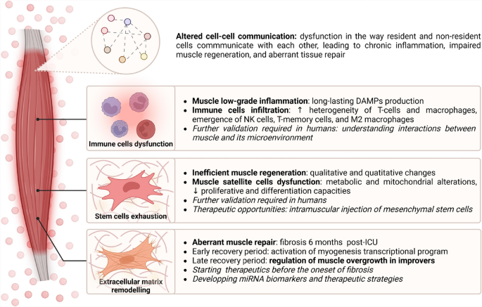
Muscle inflammation can also be related to immune cell infiltration. Nakanishi et al. demonstrated that infiltration of neutrophils in muscles led to muscle weakness in sepsis-surviving mice [133]. Dos Santos et al. found macrophage infiltration in human ICU survivors 7 days after discharge, but which were no longer present at 6 months [79]. Nevertheless, macrophage infiltration has been shown to persist for up to 9 months in COVID-19 ICU survivors [84]. The communication between cells is also of primary importance in maintaining the homeostasis and function of tissue. Interactions between mononuclear cells and signals from immune and mesenchymal interstitial cells are crucial for muscle repair. Disruptions in this network can lead to chronic muscle disorders, resulting in muscle inflammation, fibrosis, adipose infiltration, and muscle atrophy [134]. However, little is known on the subject in the field of critically ill patients. To date, only one single-cell RNA sequencing study has been conducted on the skeletal muscle of surviving mice one month after sepsis, and none have been published to our knowledge on human ICU survivors. The cellular composition of the muscle microenvironment elicits a unique signature with new cell populations. Quantitative analyses of muscle cell populations reveal reduced endothelial cells, fibroblasts, and myogenic cells, alongside increased dendritic cells, neutrophils, and T-cell/macrophage heterogeneity, with emergence of NK cells, T-memory cells, and M2 macrophages [77]. Cell–cell communication was significantly altered, with enhanced signaling toward neutrophils and reduced toward fibroblasts [135]. Inflammation-related pathways were highly dysregulated, notably with strong upregulation of DAMP-associated genes in multiple cell types. Collectively, these data implicate fibroblasts, endothelial cells, neutrophils, T-cells, and DAMP-related signaling in skeletal muscle microenvironment remodeling post-sepsis, likely reflecting altered intercellular communication that may promote chronic inflammation and impaired repair. These findings require validation in humans, particularly regarding immune cell landscape and intercellular communication in skeletal muscle of ICU survivors. Understanding the interactions between muscle cells and their microenvironment seems crucial for elucidating the mechanisms underlying sustained low-grade inflammation and its impact on physical functioning [136] (Fig. 5 and Table 1).
Biological mechanisms underlying post-ICU muscle weakness at the cellular and molecular levels: alteration of intercellular communication. Further research directions are highlighted in italics. DAMPs: damage-associated molecular patterns, NK cells: natural killer cells, miRNA: micro-RNAFull size image
Aberrant muscle repair and extracellular matrix remodelling
Following acute tissue injury, inflammatory cells, and stem cells coordinate to restore tissue homeostasis depending on local and systemic signals [137]. However, it fails in some conditions, leading to excessive extracellular matrix deposition and ultimately fibrosis, which impacts muscle contraction. Muscle fibrosis has been described in sepsis-surviving mice [107] and human ICU survivors 6 months after discharge [103]. Walsh et al. identified a network of genes involved in the extracellular matrix remodelling that was inversely correlated with muscle strength, and distinct miRNA signatures between 7 days and 6 months post-ICU discharge [103, 104]. Interestingly, Walsh et al. identified different miRNA profiles between ICU survivors with recovered muscle mass (improvers) and those with persistent muscle atrophy at 6 months (non-improvers). Improvers exhibited increased expression of two key miRNA regulators known to negatively regulate myoblast activation, compared with non-improvers, suggesting that these miRNAs may play a role in preventing excessive muscle growth. Overall, Walsh et al. provide strong data that aberrant muscle repair and extracellular matrix remodelling influence muscle weakness after critical illness. A better understanding of the miRNome of ICU survivors could provide the basis for developing miRNA therapeutic strategies in the future. Since patients reached a muscle function plateau at 6 months post-ICU discharge [7], these results also underline that interventions aiming to improve muscle weakness should likely start in the early convalescence phase, before the onset of fibrosis (Fig. 5 and Table 1).
Impaired regenerative capacities
Maintaining regenerative capacity after an injury is fundamental as it enables the restoration of muscle homeostasis through the activation, proliferation, and differentiation of muscle satellite cells (MuSCs). Human ICU survivors exhibited reduced satellite cell content and dysregulated muscle regeneration pathways 6 months after ICU discharge [79, 103]. Sepsis-surviving mice showed persistent necrosis and fibrosis after muscle injury, linked to MuSC metabolic and mitochondrial dysfunction impairing regeneration [138]. Post-sepsis MuSCs exhibited reduced proliferation and differentiation capacities in vitro. Additionally, RNA-seq analysis revealed a mitochondrial defect signature in MuSCs 28 days post-sepsis [139]. In conclusion, both quantitative and qualitative changes alter MuSCs homeostasis in the recovery phase, leading to impaired muscle regeneration. Mesenchymal stem cells (MSCs) of the skeletal muscle are multipotent stromal cells residing in muscle tissue that can differentiate into various cell types, playing key roles in driving muscle repair [140]. Rocheteau et al. successfully treated sepsis-surviving mice with intramuscular injection of MSCs [138], restoring muscle mass and function by improving the muscle regeneration capacities. Thus, MSCs grafting appears to be a promising therapy for ICU patients suffering from physical disabilities. However, the lack of data on the functionality of human MuSCs in the post-ICU period currently limits the feasibility of conducting randomized controlled clinical trials for this therapy (Fig. 5 and Table 1).
Management of post-ICU muscle weakness
Preventive approaches
Sarcopenia represents the main modifiable component of pre-ICU health status, and addressing this condition may contribute to improving long-term physical outcomes among ICU survivors. Several modifiable risk factors, including low physical activity levels, sedentary behaviour, and inadequate nutritional intake, have been identified as key contributors to sarcopenia and should be systematically screened for [24, 141]. Once sarcopenia is confirmed, targeted interventions combining resistance exercise, nutritional optimization (particularly sufficient protein intake of 1 to 1.5 g/kg/day), and reduction of sedentary behaviour have demonstrated efficacy in preventing or mitigating adverse health outcomes [24, 141, 142] (Table 2).
Table 2 Overview of current evidence on clinical risk factors and management strategies for muscle weakness across different phases of critical illness (pre-ICU, ICU stay, and post-ICU), along with identified gaps and directions for future research
Curative approaches
General management
Implementing evidence-based approaches to minimize the duration of ICU stay appears essential in preventing long-term physical disabilities in ICU survivors. One of the most widely recommended strategies is the ABCDEF bundle, a multimodal, patient-centred approach designed to reduce ICU-acquired complications. The bundle includes: A — Assess, prevent, and manage pain; B — Both spontaneous awakening and breathing trials; C — Choice of analgesia and sedation; D — Delirium assessment and management; E — Early mobility and exercise, and; F — Family engagement and empowerment. Adherence to the ABCDEF bundle is linked to shorter ICU stay, less delirium, reduced ventilation duration, and better recovery, with a dose–response relationship indicating greater benefits with higher compliance [143,144,145] (Table 2).
Physical therapy
Although widely studied in critically ill patients, physical therapy shows limited evidence of long-term functional benefit. (1) Despite heterogeneity in results, the overall effect of studies assessing protocolized early and progressive mobilization within the ICU is favourable regarding short-term physical outcomes (e.g. muscle strength at ICU discharge, mechanical ventilation duration and length of stay). While the benefits of early active mobilization are transient and appear not to affect long-term outcomes (e.g. physical recovery or quality of life) [146,147,148,149,150,151,152,153,154,155,156], current guidelines strongly recommend starting the early mobilization, “adapted to the patient’s resilience and general condition “, within the first days in the ICU [157]. (2) Evoked muscle activation using neuromuscular electrical stimulation (NMES) does not improve muscle strength, but increases muscle mass by up-regulating Myhc gene expression. No benefit has been observed on long-term outcomes [155, 158,159,160]. (3) Functional physical therapy, such as in-bed cycling or functional electrical stimulation (FESCE), is currently thought not to improve muscle strength, muscle mass, or physical outcomes in the short- or long-term, however they are safe [155, 161,162,163]. (4) Few studies have assessed exercise training post-ICU, and current evidence does not support a clear benefit for physical recovery [164]. Walsh et al. conducted the first RCT assessing combined physical and nutritional rehabilitation during the post-ICU hospital ward, but it showed no improvement in physical recovery outcomes [165] (Table 2).
To explain these results, we could put forward some hypotheses: (a) In a recent negative study evaluating FESCE in critically ill patients receiving mechanical ventilation, RNA sequencing of electrically stimulated versus non-stimulated muscle tissue revealed that the intervention's inefficacy was likely due to persistent inflammatory and metabolic dysregulation [161, 166]; (b) Since physical fitness improves physiologically with exercise training over several weeks, it is possible that the dose and/or duration of the interventions were not sufficient to observe long-term results; (c) the exercise training response is highly heterogeneous among individuals (e.g. in elderly, diabetic, or athletic populations), with some subjects experiencing greater improvements than others [167, 168]. Further research should specify the optimal dose (duration, intensity, frequency, and volume) of early mobilization and the role of evoked muscle activation strategies (e.g. NMES) in the physical rehabilitation arsenal. Additionally, studies should investigate the effectiveness of a structured training program, i.e. intervention in which patients are engaged in planned (over at least 12 weeks), time-adaptive, individualized, supervised exercise programs [169]. Increasing the cumulative dose of exercise from the ICU stay through post-ICU convalescence may contribute to improved long-term physical outcomes. However, the optimal type of exercise (e.g., aerobic, resistance, balance) and its appropriate dose remains to be determined (Table 2).
Nutritional approaches
Following ICU discharge, nutritional intake is generally considered to be insufficient, especially for those who had their enteral nutrition stopped prematurely in the ICU ward and depended thereafter solely on an oral diet [170]. Nevertheless, reaching the protein calorie target, based on formula-based calculations by dieticians, was shown not to be associated with improvement in recovery [171]. Protocol-guided and individualised nutritional support by specialist dietitians has proven its effectiveness among non-critically ill medical patients by lowering mortality [172]. Such a strategy could be applied to survivors after ICU discharge. In addition, as swallowing disorders are common post-ICU, early detection is essential to allow timely adaptation of dietary texture and initiation of compensatory strategies (e.g., supraglottic swallowing). Pharyngeal electrical stimulation via gastric feeding tube shows promise, but requires further validation [45, 46] (Table 2).
Conclusion
After an initial improvement in the first months after critical illness, muscle weakness persists for years and impacts the long-term prognosis of survivors, increasing healthcare facilities and socio-economic costs. Pre-ICU health status, ICU-related conditions and treatments, and post-ICU management all influence the physical recovery. The pathophysiology of post-ICU muscle weakness is dynamic over time, from the ICU stay through post-ICU convalescence, with some mechanisms fading (e.g. anabolic resistance) while others arise (e.g. impaired regeneration) or persist (e.g. mitochondrial dysfunction). It involves complex dysfunctions spanning the CNS to skeletal muscle, and implies multifaced and interconnected cellular, molecular, and systemic processes. However, these mechanisms remain poorly understood, warranting further investigation in humans through advanced biotechnological tools to uncover novel biological therapeutic strategies. Physical rehabilitation within ICU improves short-term outcomes but its long-term benefits appear limited, potentially due to insufficient cumulative dose, persistent inflammatory and metabolic disturbances in skeletal muscle, and inter-individual variability in exercise response. To date, nutrition- and rehabilitation-based strategies following ICU discharge have not demonstrated evidence of a benefit, but this remains an emerging field and evidence from other applications indicates it warrants further investigation [164]. Interventions targeting post-ICU muscle weakness should commence in the early convalescence phase, before the onset of fibrosis. Multi-approach treatments combining nutritional, physical, and biological approaches during and after the ICU stay should be considered to improve long-term physical outcomes.
ARDS: Acute respiratory distress syndrome CCI: Chronic critical Illness CNS: Central nervous system CLP: Cecal ligation puncture DAMPs: Damage-associated molecular patterns EMG: Electromyography ICU: Intensive care unit ICUAW: ICU-acquired weakness IGFBP3: Insulin-like growth factor binding protein 3 ME/CFS: Myalgic encephalomyelitis/chronic fatigue syndrome miRNA: Micro-RNA MRC: Medical research council MSCs: Mesenchymal stem cells MuSCs: Muscle satellite cells NMBA: Neuromuscular blocking agents NMJ: Neuromuscular junction PICS: Post-intensive care syndrome PICU: Pediatric ICU PNS: Peripheral nervous system QOL: Quality of life RCT: Randomized controlled trial rT3: Reverse triiodothyronine SASP: Senescence-associated secretory phenotype UPS: Ubiquitin-proteasome system VO2: Systemic oxygen consumption
Article CAS PubMed Google Scholar
Article PubMed PubMed Central Google Scholar
Article PubMed PubMed Central Google Scholar
Article PubMed PubMed Central Google Scholar
Article PubMed PubMed Central Google Scholar
Article PubMed PubMed Central Google Scholar
Article PubMed PubMed Central Google Scholar
Article PubMed PubMed Central Google Scholar
Article CAS PubMed Google Scholar
Article PubMed Google Scholar.%20A%20randomized%20controlled%20trial&journal=Am%20J%20Respir%20Crit%20Care%20Med&doi=10.1164%2Frccm.201909-1810OC&volume=201&issue=7&pages=814-822&publication_year=2020&author=Deane%2CAM&author=Little%2CL&author=Bellomo%2CR&author=Chapman%2CMJ&author=Davies%2CAR&author=Ferrie%2CS)
Article PubMed Google Scholar70183-8&volume=1&issue=8&pages=621-629&publication_year=2013&author=Hermans%2CG&author=Casaer%2CMP&author=Clerckx%2CB&author=G%C3%BCiza%2CF&author=Vanhullebusch%2CT&author=Derde%2CS)
Article CAS PubMed Google Scholar
Article CAS PubMed Google Scholar
Article PubMed PubMed Central Google Scholar
Article PubMed PubMed Central Google Scholar
Article PubMed PubMed Central Google Scholar
Article PubMed PubMed Central Google Scholar
Article CAS PubMed Google Scholar
Article PubMed PubMed Central Google Scholar
Article CAS PubMed Google Scholar
Article CAS PubMed Google Scholar
Article CAS PubMed Google Scholar&doi=10.1152%2Fjapplphysiol.00852.2010&volume=110&issue=1&pages=109-115&publication_year=2011&author=Greising%2CSM&author=Baltgalvis%2CKA&author=Kosir%2CAM&author=Moran%2CAL&author=Warren%2CGL&author=Lowe%2CDA)
Article CAS PubMed Google Scholar
Article PubMed PubMed Central Google Scholar
Article PubMed PubMed Central Google Scholar
Article PubMed PubMed Central Google Scholar
Article CAS PubMed Google Scholar
Article CAS PubMed Google Scholar
Article CAS PubMed Google Scholar
Article PubMed PubMed Central Google Scholar
Article PubMed PubMed Central Google Scholar
Article Google Scholar90823-1&volume=322&issue=8365%E2%80%938366&publication_year=1983&author=Gasparetto%2CA&author=Corbucci%2CGG&author=Candiani%2CA&author=Gohil%2CK&author=Edwards%2CRHT)
Article PubMed PubMed Central Google Scholar
Article CAS PubMed Google Scholar09459-X&volume=360&issue=9328&pages=219-223&publication_year=2002&author=Brealey%2CD&author=Brand%2CM&author=Hargreaves%2CI&author=Heales%2CS&author=Land%2CJ&author=Smolenski%2CR)
Article PubMed PubMed Central Google Scholar
Article PubMed PubMed Central Google Scholar
Article PubMed PubMed Central Google Scholar
Article CAS PubMed Google Scholar
Article PubMed PubMed Central Google Scholar
Article CAS PubMed Google Scholar
Article CAS PubMed Google Scholar
Article CAS PubMed Google Scholar
Article CAS PubMed Google Scholar
Article PubMed PubMed Central Google Scholar
Article PubMed PubMed Central Google Scholar
Article PubMed PubMed Central Google Scholar
Article PubMed PubMed Central Google Scholar
Article PubMed PubMed Central Google Scholar
Article PubMed PubMed Central Google Scholar
Article PubMed PubMed Central Google Scholar
Article CAS PubMed Google Scholar
Article Google Scholar60658-9&volume=373&issue=9678&pages=1874-1882&publication_year=2009&author=Schweickert%2CWD&author=Pohlman%2CMC&author=Pohlman%2CAS&author=Nigos%2CC&author=Pawlik%2CAJ&author=Esbrook%2CCL)
Article PubMed PubMed Central Google Scholar
Article PubMed Google Scholar%3A%20a%20multicentre%2C%20parallel-group%2C%20randomised%20controlled%20trial&journal=Thorax&doi=10.1136%2Fthoraxjnl-2016-209858&volume=73&issue=3&pages=213-221&publication_year=2018&author=Wright%2CSE&author=Thomas%2CK&author=Watson%2CG&author=Baker%2CC&author=Bryant%2CA&author=Chadwick%2CTJ)
Article PubMed PubMed Central Google Scholar
Article PubMed PubMed Central Google Scholar
Article PubMed PubMed Central Google Scholar
Article PubMed PubMed Central Google Scholar
Article CAS PubMed Google Scholar
Article CAS PubMed Google Scholar
Article PubMed Google Scholar32776-4&volume=393&issue=10188&pages=2312-2321&publication_year=2019&author=Schuetz%2CP&author=Fehr%2CR&author=Baechli%2CV&author=Geiser%2CM&author=Deiss%2CM&author=Gomes%2CF)
Not applicable
The study was supported by the French National Research Agency (ANR-23-CE14-0063–01). AP was funded by the French Ministry for Research and supported by a Ph.D. scholarship from Lille University. RR was funded by a Ph.D. scholarship from Lille University. CB was funded by Lille University for a Master’s degree scholarship.
Authors and Affiliations
A.P. wrote the main manuscript, and all authors provided substantial review and revisions.
Correspondence to Alexandre Pierre.
Springer Nature remains neutral with regard to jurisdictional claims in published maps and institutional affiliations.
Open Access This article is licensed under a Creative Commons Attribution 4.0 International License, which permits use, sharing, adaptation, distribution and reproduction in any medium or format, as long as you give appropriate credit to the original author(s) and the source, provide a link to the Creative Commons licence, and indicate if changes were made. The images or other third party material in this article are included in the article's Creative Commons licence, unless indicated otherwise in a credit line to the material. If material is not included in the article's Creative Commons licence and your intended use is not permitted by statutory regulation or exceeds the permitted use, you will need to obtain permission directly from the copyright holder. To view a copy of this licence, visit http://creativecommons.org/licenses/by/4.0/.