Técnicas para otimizar a punção arterial radial guiada por U
Este artigo apresenta técnicas avançadas para otimizar a punção arterial radial guiada por ultrassom, destacando quatro aspectos principais que melhoram as taxas de sucesso do procedimento. A cateterização guiada por ultrassom aumenta a taxa de sucesso em 20-30% e reduz complicações em 40%. As técnicas incluem a relação plano-agulha, modificações no probe de ultrassom, abordagens relacionadas à ar
Visão geral
RESUMO EM PORTUGUÊS
Resumo Detalhado do Conteúdo
Este artigo apresenta técnicas avançadas para otimizar a punção arterial radial guiada por ultrassom, focando em quatro aspectos principais que podem melhorar significativamente as taxas de sucesso do procedimento.
📊 Contexto e Importância
A cateterização da artéria radial guiada por ultrassom tornou-se uma técnica rotineira em UTIs, departamentos de emergência e salas cirúrgicas. Comparada à palpação tradicional, a orientação por ultrassom:
No entanto, a técnica ainda pode ser aprimorada em populações especiais: pacientes pediátricos, idosos, obesos, hipotensos e com doença renal crônica.
🔬 Metodologia da Revisão
Os autores realizaram uma busca abrangente nas bases de dados PubMed e MEDLINE, analisando artigos publicados entre outubro de 1992 e abril de 2025. Foram selecionados 49 artigos de pesquisa, principalmente ensaios clínicos randomizados, focados em técnicas que facilitam o sucesso da canulação.
🎯 Quatro Aspectos Principais para Otimização
O artigo organiza as técnicas em quatro categorias fundamentais, ilustradas nas figuras originais:
1️⃣ Relação Plano-Agulha
Técnica DNTP (Dynamic Needle Tip Positioning)
Com visualização em eixo curto fora do plano, a agulha perfura a pele em 30° a 40° até a ponta hiperecóica aparecer na imagem. O probe é movido proximalmente até a ponta desaparecer, e a agulha é avançada alguns milímetros até reaparecer. Este processo gradual é repetido até que pelo menos 1 cm da agulha esteja dentro do lúmen arterial.
Técnica LAX-IP combinada com SAX-OOP
Localiza-se a artéria no meio da imagem no plano de eixo curto, depois o probe é rotacionado 90° paralelo ao eixo longo da artéria quando a agulha perfura a pele. Uma vez que a ponta aparece dentro da artéria, o probe é rotacionado novamente 90° para o eixo curto.
Abordagem Oblíqua (OA-IP)
O probe é rotacionado 15° com o eixo longitudinal cruzando obliquamente a artéria radial. Isso elimina o artefato de espessura de seção, onde a ponta da agulha está dentro da largura de elevação do feixe ultrassônico mas fora do plano de varredura, proporcionando maior precisão.
2️⃣ Modificações no Probe de Ultrassom
Linha de Desenvolvimento (Developing Line)
Um fio amarrado no ponto médio do probe cria uma sombra de baixa densidade óbvia na imagem ultrassonográfica. Linhas duplas paralelas ao redor do ponto médio também podem ser usadas como marcas guia.
Probe Escalonado (Scaled Probe)
O probe é marcado com intervalos de 2 mm: lado esquerdo marcado como -5, -4, -3, -2, -1 e lado direito como 1, 2, 3, 4, 5, com ponto médio 0. Isso localiza com precisão a artéria e ajuda a encontrar a ponta da agulha mais rapidamente.
Probe Tipo T (T-type Probe)
Permite observação simultânea de imagens LAX-IP e SAX-OOP. A visualização transversal localiza a artéria enquanto a longitudinal direciona a canulação, sem necessidade de mover o probe.
Sistema Assistido por Laser
A orientação por laser projeta o caminho da artéria na pele, fornecendo direção mais precisa de inserção tanto nas abordagens LAX-IP quanto SAX-OOP.
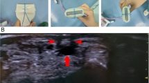
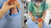
Displays Montados na Cabeça (HMDs - Smart Glasses)
A imagem de ultrassom em tempo real é exibida na parte superior dos óculos inteligentes, enquanto o campo de trabalho (local de canulação) é visível na parte inferior. O operador pode focar simultaneamente na tela de ultrassom e no campo do procedimento sem mover a cabeça, melhorando a taxa de sucesso na primeira tentativa.
3️⃣ Técnicas Relacionadas à Artéria do Paciente
Compressão Arterial
A compressão da artéria radial distal aumenta a área transversal. A oclusão da artéria ulnar 1-2 cm proximal à prega distal do punho também aumenta o diâmetro arterial. No entanto, há resultados conflitantes na literatura quanto à eficácia universal desta técnica.
Torniquete Distal
Inspirado pelo torniquete proximal para punção venosa periférica, o torniquete distal ao processo estiloide ulnar pode inflar a artéria radial proximal e facilitar a canulação tanto com palpação quanto com orientação ultrassonográfica.
Agentes Subcutâneos
Nitroglicerina: Meta-análise demonstrou que nitroglicerina tópica ou subcutânea facilita o acesso à artéria radial e diminui complicações locais. Em estudo pediátrico com 113 crianças, injeção subcutânea de nitroglicerina (5 μg/kg em 0,5 ml) aumentou o diâmetro arterial e melhorou a taxa de sucesso na primeira tentativa.
Lidocaína: Além da anestesia local, a injeção de 0,2-0,5 ml de lidocaína 1% na camada subcutânea cria uma pápula cutânea. O probe posicionado contra a inclinação proximal elimina o espaço entre pele e probe, permitindo inclinação de aproximadamente 90° e melhorando a visualização.
Solução Salina: Para artérias localizadas < 2 mm abaixo da superfície da pele, a injeção subcutânea de salina (< 2 ml) entre a pele e a artéria produz uma área anecóica, aumentando a profundidade para 2-4 mm. Isso melhora os sinais ultrassonográficos e a visibilidade da artéria e ponta da agulha.
Bloqueio do Nervo Mediano
Bloqueio do nervo mediano guiado por ultrassom no antebraço médio usando 5 ml de bupivacaína 0,5% causa vasodilatação arterial e aumento da velocidade do fluxo sanguíneo da artéria radial. Estudo randomizado com 92 gestantes mostrou que o bloqueio com 5 ml de lidocaína 0,5% aumentou a taxa de sucesso na primeira tentativa após falha na canulação.
Aquecimento Palmar
Aumentar a temperatura da palma dilata os vasos. Aquecer o local da artéria radial por três minutos com a palma (manobra de Balbay) diminuiu as tentativas de punção. Estudos voluntários revelaram que a área transversal da artéria radial mostra tendência crescente ao longo do tempo com aquecimento.
Seleção do Local de Inserção
Locais ótimos para a técnica DNTP incluem:
4️⃣ Técnicas Relacionadas à Agulha de Inserção
O artigo menciona técnicas específicas relacionadas à agulha de inserção que podem otimizar o procedimento, conforme ilustrado na figura correspondente.
💡 Considerações Práticas
As técnicas são apresentadas considerando três fatores importantes:
🎓 Conclusão
Este artigo fornece uma revisão abrangente e prática de técnicas inovadoras para melhorar o sucesso da punção arterial radial guiada por ultrassom. A estratificação em quatro aspectos modificáveis - relação plano-agulha, probe ultrassonográfico, artéria do paciente e agulha de inserção - oferece aos profissionais múltiplas opções para otimizar o procedimento em diferentes cenários clínicos e populações de pacientes.
As evidências apresentadas demonstram que a combinação apropriada dessas técnicas pode aumentar significativamente as taxas de sucesso, especialmente em populações desafiadoras, enquanto reduz complicações e o número de tentativas necessárias.
Abstract
Ultrasound-guidance increases success rate and reduces complications compared to traditional palpation for radial artery catheterization. However, in special groups of patients, such as pediatric, older, obese, hypotensive population, and patients with chronic kidney disease (CKD), ultrasound-guided arterial cannulation can still be improved. The current available evidence revealed some techniques for improvement of success rate, in consideration of various modifiable factors during the procedure. In this narrative review, we summarized and illustrated techniques in four different aspects related to plane-needle relationship, ultrasound probe, patient’s artery, and insertion needle, which may provide inspiration for better practice in the future.
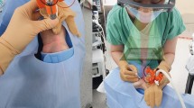
Background
With broad application of ultrasonography in clinical practice, ultrasound-guided radial artery catheterization has become a routine technique for patients in the intensive care unit, emergency department, and operating room [1]. Full evidence has shown that compared to traditional palpation, ultrasound guidance increases the success rate by 20–30% [2,3,4,5]. The incidences of both major complications (0.18%, pseudoaneurysm requiring intervention and permanent artery occlusion) and minor complications (14.93%, hematoma formation and bleeding at the puncture site) decrease by 40% [6,7,8,9,10]. However, techniques of ultrasound-guided arterial cannulation can still be improved in special groups, such as pediatric, older, obese, hypotensive, and CKD patients. The past decade witnessed the development of various useful techniques for better practice in arterial catheterization. The increase in success rates depends mainly on the modification of the plane-needle relationship, ultrasound probe, patient’s artery, and insertion needle. The goal of this narrative review is to summarize techniques in these four aspects for improvements in the success rate of ultrasound-guided radial artery catheterization.
Methods
Search strategy
This was a qualitative non-meta-analysis narrative review. Comprehensive literature search using PubMed and MEDLINE databases was performed for articles written in English from October 1992 to April 2025. The electronic search strategy included the following keywords “Ultrasonography”[MeSH], “Radial Artery”[MeSH], “Catheterization, Peripheral”[MeSH] and a combination of these using Boolean operators “and”, “or” and ‘not’. Types of study including meta-analysis, systematic reviews, clinical trials, cohort studies, case reports, editorials and letters were considered. All obtained articles were assessed by two independent reviewers (Jiamei He and Xiaoli Liu) through careful evaluation of their methodology, sample size, study design and relevance to the topic of ultrasound-guided radial artery catheterization. In cases of disagreement, a third author (Qingyu Xiao) completed the evaluation to reach consensus.
Inclusion and exclusion criteria
For a study to be included, it had to be relevant to the topic of radial artery catheterization under ultrasound guidance, with investigation in techniques facilitating successful cannulation, from perspectives of improving success rates, reducing the number of attempts, procedure duration, or complication rates. Studies exploring other anatomical locations (dorsalis pedis artery, femoral artery, or distal radial artery) for ultrasound-guided arterial cannulation were excluded.
Results
Studies for analysis
The initial search criteria yielded 433 original articles in search of the keywords, with 136 revealing outcomes closely related to the enhancement of ultrasound-guided radial artery catheterization. A total of 49 research articles were selected for final analysis, most of which were randomized clinical trials. Details of the studies were presented in the Supplementary Table S1 (Additional file 1).
Techniques
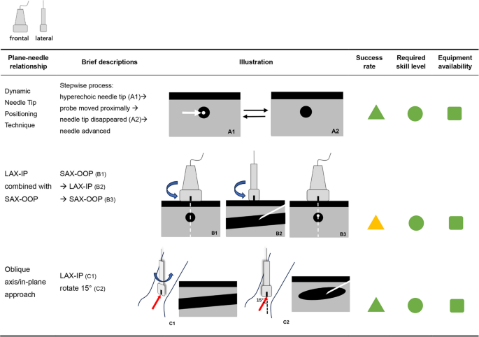
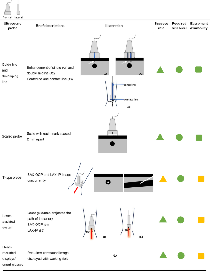
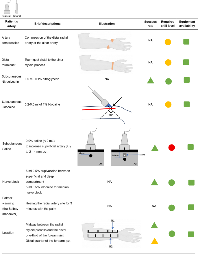
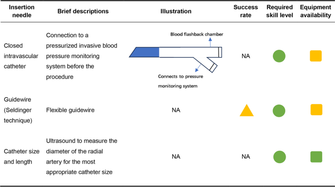
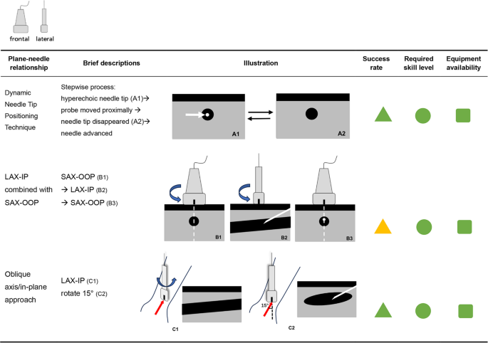
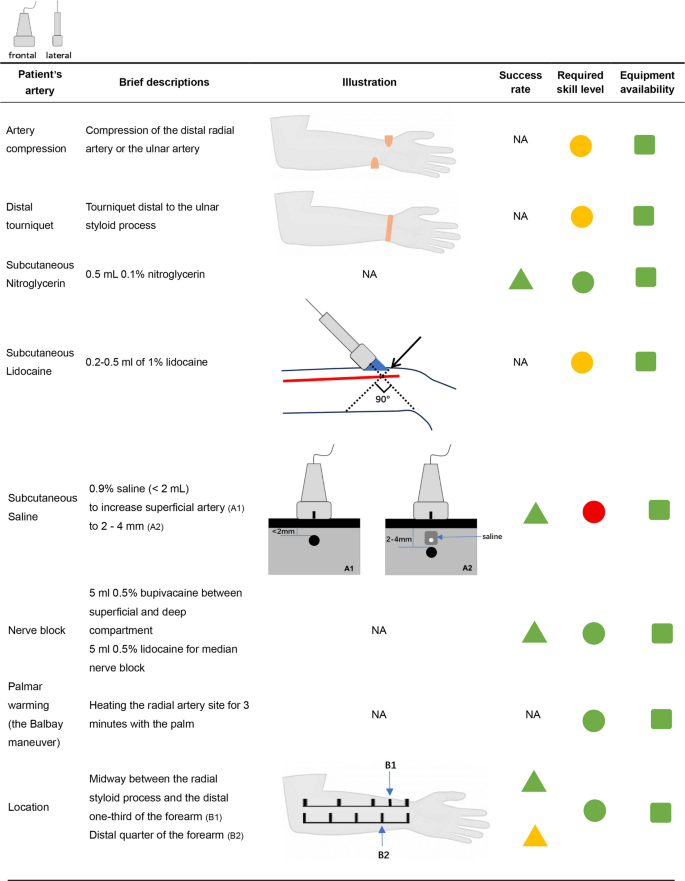
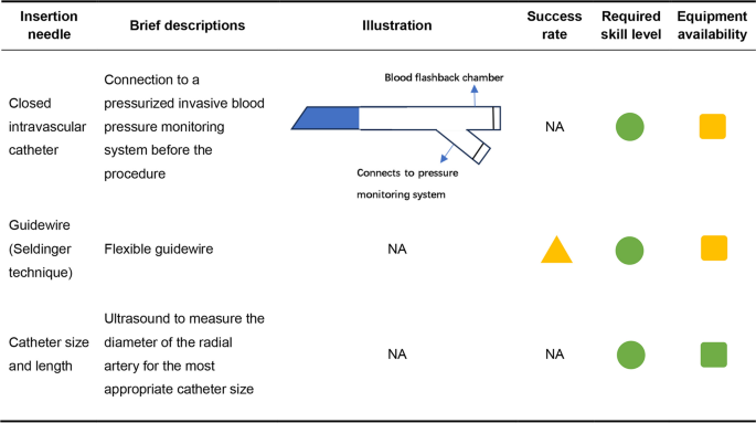
Stratification of procedural strategies in four different aspects related to plane-needle relationship, ultrasound probe, patient’s artery, and insertion needle was summarized and illustrated in Figs. 1, 2, 3, and 4. Success rates, required skill levels, and equipment availability were demonstrated in different colors (triangle: green ≥ 80%, yellow < 80%; square: green-low, yellow-moderate, red-high; circle: green-high, yellow-limited).
Techniques modifying plane-needle relationship. white arrow: needle tip; red arrow: direction of needle insertion. LAX-IP, long-axis in-plane; SAX-OOP, short-axis out-of planeFull size image
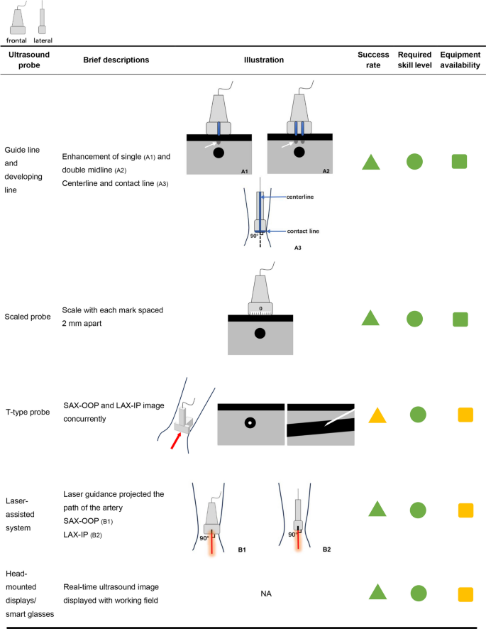
Techniques modifying the ultrasound probe white arrows: low-density shadows; red arrow: direction of needle insertion. LAX-IP, long-axis in-plane; SAX-OOP, short-axis out-of-plane; NA, not availableFull size image
Techniques related to patient’s artery. blue triangle: skin wheal created by subcutaneous lidocaine; red line: radial artery; black arrow: direction of needle insertion. NA, not availableFull size image
Techniques related to insertion needle. NA, not availableFull size image
Dynamic Needle Tip Positioning (DNTP) Technique [11]
With a short-axis out-of-plane view of the radial artery, the needle punctured the skin at 30° to 40° until the hyperechoic needle tip appeared on the ultrasound image. The ultrasound probe was moved proximally until the hyperechoic tip disappeared. The needle was advanced a few millimeters until the tip appeared again. This stepwise process was repeated until at least 1 cm of the needle was inside the arterial lumen when the catheter was eventually threaded off.
LAX-IP combined with SAX-OOP technique [12]
The radial artery was located in the middle of the ultrasound image with the short-axis plane. Then the probe was rotated 90◦ around its midpoint parallel to the long axis of the artery when the needle punctured the skin. Once the needle tip appeared inside the artery, the probe was rotated 90◦ to the short-axis plane again and adjusted until a hyperechoic highlight was found. The remaining steps are the same as DNTP technique.
Oblique axis/in-plane (OA-IP) approach [13]
In traditional LAX-IP technique, arterial wall puncture was common because of section-thickness artefact, meaning that the needle tip was within the elevation width of the ultrasonic beam but actually out of the scanning plane. For OA-IP approach, the probe was rotated 15° with the longitudinal axis obliquely crossing the radial artery. Therefore, when the needle tip was shown inside the vessel lumen of ultrasound images, the location was accurate, which accounted for the higher success rate compared with the LAX-IP approach.
Guide line and developing line
A developing line is an enhancement of the midline with a string tied at the midpoint of the ultrasound probe, making an obvious low-density shadow on the ultrasound image. Double developing lines were parallel lines guide marks around the midpoint of the ultrasound probe [14,15,16,17]. In another line-developing approach, a suture along the probe side was the centerline while the second suture at the lower edge of the probe was the contact line, which maintained the center axis of the artery in the ultrasonic plane [18].
Scaled probe
The ultrasound probe could be scaled, with each mark spaced 2 mm apart, meaning that the left side was marked as − 5, − 4, − 3, − 2, − 1 while the right side 1, 2, 3, 4, 5, and the midpoint 0. This technique accurately localized the artery, which helped operators find the tip of the needle more quickly, thus improving success rates and shortening puncture time compared to traditional ultrasound probe [19].
T-type probe
A t-type ultrasound probe allowed observation of both LAX-IP and SAX-OOP images at the same time. The cross-sectional view helped localize the artery, while the longitudinal view directed the cannulation. Practitioners could advance the needle toward the radial artery without moving the probe [20].
Laser-assisted system
The laser guidance projected the path of the artery on the skin; therefore, it provided more precise direction of insertion in both LAX-IP and SAX-OOP approaches compared with traditional ultrasound guidance [21, 22].
Head-mounted displays (HMDs)
The HMDs are smart glasses with a simultaneous ultrasound screen. The real-time ultrasound image was displayed on the upper part, while the working field, including the needle and probe, was visible through the lower part. With smart glasses, the operator can focus on the ultrasound screen and procedure field (cannulation site) simultaneously without moving the head, which improved the first attempt success rate compared to the routine ultrasound guidance group [23,24,25].
Artery compression
In previous research, compression of the distal radial artery increased the cross-sectional area. Occlusion of the ulnar artery 1–2 cm proximal to the distal wrist crease also increased arterial diameter [26]. However, there were conflicting results. A volunteer study showed that ulnar artery compression did not influence the diameter of the ipsilateral radial artery [27]. Another study revealed that neither compression of the distal radial nor the ulnar artery increased the size of the radial artery in patients with previous or current peripheral vascular procedures [28]. These articles only investigated the diameter changes and did not apply the technique to arterial cannulation, which could be further studied.
Distal tourniquet
Distal tourniquet was inspired by proximal tourniquet for peripheral venipuncture. Studies found that tourniquet distal to the ulnar styloid process could inflate the proximal radial artery and facilitate radial artery cannulation with both palpation and ultrasound guidance [29, 30].
Subcutaneous agent s
Nitroglycerin
An updated meta-analysis demonstrated that topical or subcutaneous nitroglycerin has the potential to facilitate radial artery access and decrease local complications [31]. A pediatric study of 113 children also found that subcutaneous nitroglycerin injection (5 μg/kg in 0.5 ml) before cannulation increased the diameter, improved the first-attempt success rate, and reduced the overall complication rates compared with the normal saline group [32].
Lidocaine
Subcutaneous administration of lidocaine is for routine local anesthesia. Anesthesiologists also found the mechanical effect of lidocaine during cannulation. They injected 0.2–0.5 ml of 1% lidocaine into the subcutaneous layer to create a skin wheal. The probe was positioned against the proximal slope of the skin wheal, which eliminated the gap between the skin and the probe, allowing the probe to be tilted approximately 90° to the needle and enhancing visualization of both the artery and the needle [33]. A randomized clinical trial is required for a higher level of evidence.
Saline
DNTP technique was challenging in superficial target vessels because needle tip control was difficult at shallow depths immediately after skin perforation [34, 35]. Injection of saline between the skin and the artery produced an anechoic area, enhancing the ultrasound signals and improving the visibility of the artery and needle tip. For arteries located < 2 mm below the skin surface, increasing the depth to 2 to 4 mm by subcutaneous saline injection (< 2 ml) reduced catheterization time and improved the success rate [36].
Nerve block
Ultrasound guided median nerve block using 5 ml 0.5% bupivacaine in the mid forearm between the superficial and deep compartments caused arterial vasodilation, and an increase in radial artery blood flow velocity [37]. A randomized controlled clinical trial recruiting 92 pregnant women found that ultrasound guided median nerve block with 5 ml of 0.5% lidocaine increased the first attempt success rate following failed radial artery cannulation [38].
Palmar warming
It seems common sense that increasing the temperature of the palm can dilate the vessels. A volunteer study revealed that the radial artery cross-sectional area showed an increasing trend over time [39]. Heating the radial artery site for three minutes with the palm (the Balbay maneuver) decreased puncture attempts [40]. The effect of palmar warming on real-time ultrasound practice requires more clinical studies.
Insertion site selection
Midway between the radial styloid process and the distal one-third of the forearm may be optimal for the DNTP technique [41]. Also, the distal quarter of the forearm might also be a better option compared with the midpoint of the forearm [42]. At least 4–6 cm from the wrist crease area or styloid process was also reported as a choice [43,44,45]. Ultrasound scanning of radial arteries prior to puncture to find an optimal site with a larger cross-sectional area was associated with a significant increase in the radial access success rate and a reduction in duration, which was simple but effective [46, 47].
Closed intravascular catheter
The side port of a 20-G closed intravascular catheter kit was connected to a pressurized invasive blood pressure monitoring system before the procedure, which improved the success rate of catheterization because intra-catheter real-time pressure could be observed continuously; also, continuous flush fluid from the pressure monitoring system enhances needle tip echogenicity [48].
Guidewire (Seldinger technique)
With the help of both guidewire and ultrasound localization before the procedure, the success rate of cannulation improved significantly in challenging cases [49, 50]. Whether the Seldinger technique could facilitate real-time catheterization under ultrasound guidance requires more investigation.
Catheter size and length
Using ultrasound to measure the diameter of the radial artery and choose the most appropriate catheter size before proceeding was helpful in avoiding complications [51]. For extremely small artery sizes measured by ultrasound just before puncture, smaller catheters or even venous cannulas may be helpful in increasing success rates.
Discussion
Application of ultrasound significantly increases the successful rate of radial artery catheterization, even for inexperienced residents or trainees [11, 16, 22, 52, 53], because the ultrasound image provides a visible tool for the procedure. In traditional points of view, pediatric, older, obese, hypotensive, and CKD patients are the most challenging groups for catheterization due to the weak pulses. Ultrasound has demonstrated unequivocal efficacy and safety with the procedure, improving first attempt success rates with children, the obese, or patients with unstable clinical vital signs and shock [54]. With the help of advanced and modified techniques, the procedure of catheterization can be more efficient, causing less harm in these special groups of patients. Success metrics of different techniques stratified by patient subgroup were summarized in Table 1.
Table 1 Success metrics of different techniques
Pediatric patients
The small diameter of the pediatric radial artery makes catheterization challenging. Previous research showed that a cross-sectional area of ≤ 1 mm2 was a predictor for failure of ultrasound-guided radial artery catheterization at the first attempt among children [46]. Arterial spasm was the most common complication for pediatric arterial insertion (4%), which added to the difficulty during the procedure [55]. Techniques for improvement of the success rate focus on three main procedures: localization, puncture, and catheter placement. A single or double developing line on the ultrasound probe contributes to accurate localization of the tiny artery by fixing the hypoechoic vessel in the midline of the ultrasound image [15]. Subcutaneous vasodilators like phentolamine and nitroglycerin increase the diameter and cross-sectional area of the artery, which may increase the success rate of the first puncture [32, 56]. Cannulation is the step that accounts for most of the failures. Threading of the catheter right inside the lumen of the artery requires accurate and confirmed direction. DNTP technique [57, 58], LAX-IP combined with SAX-OOP [12], developing line [14, 16, 52, 53] scaled probe [19], laser guidance [21] and smart glasses [23, 24] may help the operators to avoid needle redirection along the arterial line. Subcutaneous saline increases the depth of the superficial artery to an optimal depth of 2 to 4 mm, thus allowing a wider angle of needle mobility while keeping the tip in the center of the small artery [36].
Older patients
Older population is characterized by tortuous peripheral arteries [59]. Previous study showed that DNTP technique has better efficiency and safety than the LAX-IP technique for radial artery catheterization in the population aged 75 years or older because it is less affected by arterial angle tortuosity with the direction of the needle constantly modified in the lumen of the radial artery [60]. Also, arteries of the older population are less well distended due to calcification of the vessel wall with segmental stenosis [59]. Arteries could further collapse resulting from low mean arterial pressure following administration of general anesthetics. A tightened distal tourniquet increased the overall success rates of ultrasound-guided radial artery cannulation in a subgroup of patients aging 65 years or above through expansion of the cross-sectional up-forward diameter and the inner cross-over area [30].
Obese patients
Radial arteries of obese patients are usually buried in depth under thick subcutaneous adipose tissue beyond optimal depth [36]. A previous study has confirmed that obese patients with higher BMI had greater skin-to-artery distances of the radial artery [61]. Obese patients are also predisposed to adverse vascular events after arterial catheterization, including hematoma, pseudoaneurysm, and arteriovenous fistula [61, 62]. Very few studies thoroughly evaluate ultrasound-guided arterial catheterization in the overweight population. Conventional catheterization may be difficult due to weak pulse [43]. Ultrasonography contributes greatly to the accurate localization of the artery. However, the challenge lies in the subsequent procedure of arterial puncture and catheter placement. As the needle should travel a longer distance subcutaneously before it enters the artery, it is easy for the needle to wander from the desired path to the artery [17]. Developing lines on the ultrasound probe displaying vertical low-density shadows in the ultrasound image guide the puncture and cannulation procedure continuously and precisely [17]. In addition, a catheterization kit with a guidewire may also be helpful in catheter threading in consideration of the large angle between the insertion needle and the deep artery [61].
Hypotensive patients
Conventional catheterization may be difficult due to a weak pulse from hemodynamic instability [43, 55], with a reported failure rate between 10 and 50% for patients with shock [54]. Ultrasound guidance increases the success rate in patients with hypotension and shock to as high as 80–90% [43, 63]. The radial artery in shocked patients is collapsible with a small inner diameter and unobvious rapid contraction, which can be further narrowed by the compression of the ultrasound probe, making it hard to localize the artery [64]. The distal tight tourniquet technique expands the diameter and cross-sectional area of the proximal radial artery, which benefits hypotensive patients [29, 30]. Midline guidance and local zoom also help the operators to better identify and track the weak-pulsating artery [64]. Besides, scanning the radial artery for the best insertion region and cannulation direction is recommended in clinical practice [65].
CKD patients
Chronic renal failure is one of the predictors of pathological radial artery changes, including absent pulse, decreased vasoreactivity, increased arterial stiffness, and calcifications, resulting in more complications such as pseudoaneurysm, bleeding, and radial artery occlusion, which may pose unique challenges to even the most skilled health care professional [2, 8, 10, 66,67,68,69,70,71,72,73]. Ultrasound-guided radial access had significantly higher first-pass success rates, fewer attempts, and a shorter amount of time compared with palpation guidance [2]. A randomized controlled trial containing CKD patients reveals that the DNTP technique had a higher first-attempt success rate and required less time for radial artery cannulation compared to the conventional LAX-IP technique [74]. Another study showed that dialysis patients tolerated radial access well with the help of a guidewire [72]. To avoid further deterioration of vascular condition, accurate localization of the artery and real-time tracing of the catheter are both important for successful cannulation at the first attempt.
The first attempt success rate and the time to establish the arterial line is operator dependent, and there is a learning curve to become proficient in this technique [75]. Ultrasound allows faster learning of radial artery catheterization over anatomical palpation, shortens the learning curve, especially for inexperienced operators [76]. DNTP is a favorable technique with a short learning duration for novice practitioners. One-year postgraduate interns placing fewer than 15 previous arterial lines achieved a first-pass success rate as high as 75% after a 4-h course and a 4-week emergency ultrasound rotation without additional practice or training [77]. Modifications of the ultrasound probe, including developing line, t-type probe, laser guidance, and smart glasses are also friendly to non-expert operators [16, 20, 22, 23, 52]. Guidewire effectively assisted arterial catheterization by novice nurses inexperienced with ultrasound technique [78]. However, the advantage was not apparent for experienced clinicians [23]. Experience plays a more important role in techniques modifying patients’ arteries, most of which depend on manual operation from clinicians. For anesthesiologists with more than 50 arterial catheterizations in pediatric patients, subcutaneous injection of saline resulted in a first attempt success rate of 85.0% [36], while for year 2 or 3 trainee residents, the success rate was only 27.1% [79]. Therefore, repeated training is highly required when applying techniques for the improvement of arterial condition.
This article summarizes techniques from various aspects for improvement of success rates in ultrasound-guided radial artery catheterization. However, a number of limitations should be noted. First, the study mainly focused on the proximal (ventral) radial artery but not the distal radial artery in the snuff box or other peripheral arteries like the dorsalis pedis artery. Second, some evidence was from editorials or case studies, so a high level of research is required in the future. Last but not least, some advanced devices (like laser systems, smart glasses, or t-type ultrasound probes) are not readily available in clinical practice, and some other updated techniques (like magnetic needle-tracking devices) still require further exploration. More useful techniques are worth trying for a more efficient and precise procedure.
Conclusion
Techniques from perspectives of plane-needle relationship, ultrasound probe, patient’s artery, and insertion needle may improve success rates and reduce complications for ultrasound-guided radial artery catheterization.
No datasets were generated or analyzed during the current study
None.
This work was supported by Guangdong Provincial Medical Association Clinical Research Fund—Huyou Program (grant numbers 2024HY-B4016).
JH and XL analyzed and interpreted the article data regarding ultrasound-guided radial artery catheterization. SZ and QX performed graphic the procedures and drafted the manuscript. All authors read and approved the final manuscript. JH and XL contributed equally to this work and share first authorship.
Ethics approval and consent to participate
Not applicable.
Consent for publication
Not applicable.
Competing interests
The authors declare that they have no competing interests.
Springer Nature remains neutral with regard to jurisdictional claims in published maps and institutional affiliations.
Open Access This article is licensed under a Creative Commons Attribution-NonCommercial-NoDerivatives 4.0 International License, which permits any non-commercial use, sharing, distribution and reproduction in any medium or format, as long as you give appropriate credit to the original author(s) and the source, provide a link to the Creative Commons licence, and indicate if you modified the licensed material. You do not have permission under this licence to share adapted material derived from this article or parts of it. The images or other third party material in this article are included in the article’s Creative Commons licence, unless indicated otherwise in a credit line to the material. If material is not included in the article’s Creative Commons licence and your intended use is not permitted by statutory regulation or exceeds the permitted use, you will need to obtain permission directly from the copyright holder. To view a copy of this licence, visit http://creativecommons.org/licenses/by-nc-nd/4.0/.